Subdural empyema
| Subdural empyema | |
| Classification and external resources | |

| |
|---|---|
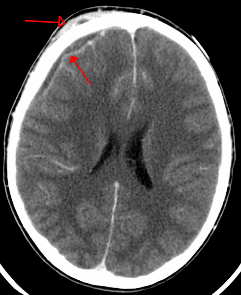
| An abscess that has let to an intracranial subdural empyema as seen on CT |
For patient information, click here
|
Subdural empyema Microchapters |
|
Diagnosis |
|
Treatment |
|
Case Studies |
|
Subdural empyema On the Web |
|
American Roentgen Ray Society Images of Subdural empyema |
Editor-In-Chief: C. Michael Gibson, M.S., M.D. [1]; João André Alves Silva, M.D. [2]
Overview
Subdural empyema, also referred to as subdural abscess, pachymeningitis interna and circumscript meningitis, is a life-threatening infection, first reported in literature approximately 100 years ago.[1] It consists of a located collection of purulent material, usually unilateral, between the dura mater and thearachnoid mater. It accounts for about 15-22% of the reported focal intracranial infections. The empyema may develop intracranially (about 95%) or in the spinal canal (about 5%), and in both cases, it constitutes a medical and neurosurgical emergency.[2] The intracranial type tends to behave like an expanding mass, causing clinical symptoms, such as fever, lethargy, headache and neurological deficits. These result from the extrinsic compression of the brain, caused not only from the inflammatory mass, but also from the inflammation of the brain and meninges. Because the subdural space has no septations, except in areas where arachnoid granulations attach to the dura mater, the subdural empyema tends to speed quickly, until it finds those boundaries. In children, subdural empyema most often happens as a complication of meningitis, while in adults it usually occurs as a complication of sinusitis, otitis media, mastoiditis, trauma or as a complication of neurological procedures.[1] The most common pathogens in the intracranial type are anaerobic and microaerophilic streptococci, but others like Escherichia coli and Bacteroides may be present simultaneously. Spinal subdural empyemas, on the other hand, are almost always caused by''streptococci'' or by staphylococcus aureus.[2] Classic clinical syndrome includes acute fever, that rapidly progresses into neurological deterioration, which if left untreated will eventually lead to a coma and death.[1] The diagnostic procedure of choice is the MRI with gadolinium enhancement. Since the clinical symptoms might be mild and unspecific initially, the rapid diagnosis and treatment are crucial. The sooner the proper treatment is initiated, the better the recovery will be. The treatment, for almost all causes, requires prompt surgical drainage and antibiotic therapy.[2] With treatment, resolution of the empyema occurs from the dural side, and, if it is complete, a thickened dura may be the only residual finding.
Historical Perspective
Pathophysiology
Causes
Differentiating Subdural empyema from other Diseases
Epidemiology and Demographics
Risk Factors
Natural History, Complications and Prognosis
Diagnosis
History and Symptoms | Physical Examination | Laboratory Findings | Electrocardiogram | X Ray | CT | MRI | Other Diagnostic Studies
Treatment
Medical Therapy | Surgery | Prevention | Cost-Effectiveness of Therapy | Future or Investigational Therapies
Case Studies
Case #1 Template:WH Template:WS
- ↑ 1.0 1.1 1.2 Agrawal, Amit; Timothy, Jake; Pandit, Lekha; Shetty, Lathika; Shetty, J.P. (2007). "A Review of Subdural Empyema and Its Management". Infectious Diseases in Clinical Practice. 15 (3): 149–153. doi:10.1097/01.idc.0000269905.67284.c7. ISSN 1056-9103.
- ↑ 2.0 2.1 2.2 Greenlee JE (2003). "Subdural Empyema". Curr Treat Options Neurol. 5 (1): 13–22. PMID 12521560.