Sandbox: dysphagia: Difference between revisions
Jump to navigation
Jump to search
Ahmed Younes (talk | contribs) No edit summary |
Ahmed Younes (talk | contribs) No edit summary |
||
| Line 1: | Line 1: | ||
{| class="wikitable" | {| class="wikitable" | ||
! rowspan="3" align="center" style="background:#4479BA; color: #FFFFFF;" |Disease | ! rowspan="3" align="center" style="background:#4479BA; color: #FFFFFF;" |Disease | ||
| Line 329: | Line 328: | ||
* Barium [[esophagogram]] | * Barium [[esophagogram]] | ||
|} | |} | ||
Revision as of 20:58, 17 November 2017
| Disease | Signs and Symptoms | Barium esophagogram | Endoscopy | Other imaging and laboratory findings | Gold Standard | |||||||
|---|---|---|---|---|---|---|---|---|---|---|---|---|
| Onset | Dysphagia | Weight loss | Heartburn | Other findings | Mental status | |||||||
| Solids | Liquids | Type | ||||||||||
| Plummer-Vinson syndrome |
|
+ | - | Non progressive | +/- | - | Normal |
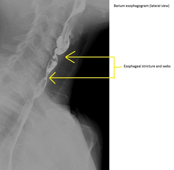 |
|
|
Triad of | |
| Esophageal stricture |
|
+ | - | Progressive | +/- | +/- | Normal |
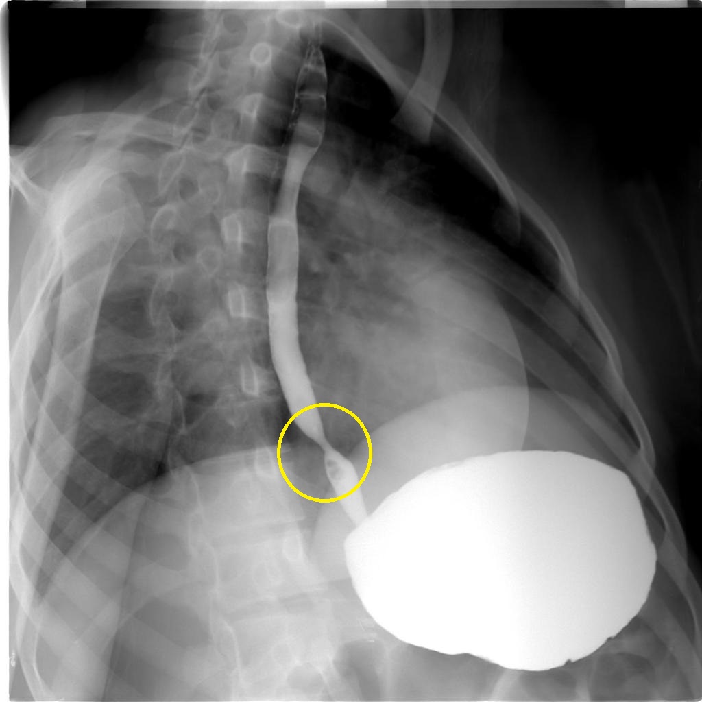 |
|
|
||
| Diffuse esophageal spasm |
|
+ | + | Non progressive | + | + | Normal |
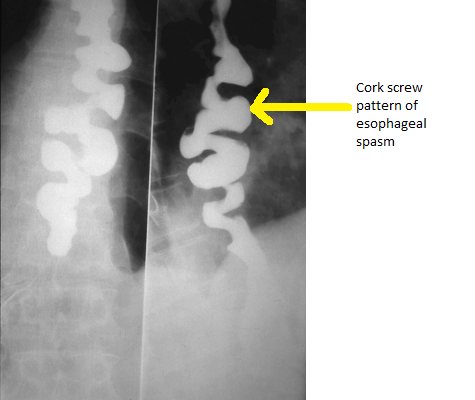 Source:By Nevit Dilmen [CC BY-SA 3.0 (https://creativecommons.org/licenses/by-sa/3.0) |
|
|
||
| Achalasia |
|
+ | + | Non progressive | +/- | - |
|
Normal |
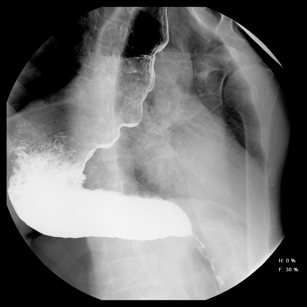 |
|||
| Systemic sclerosis |
|
+ | + | Progressive | +/- | + |
|
Normal |
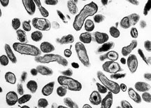 |
|
Positive serology for | |
| Zenker's diverticulum |
|
+ | - | +/- | - |
|
Normal |
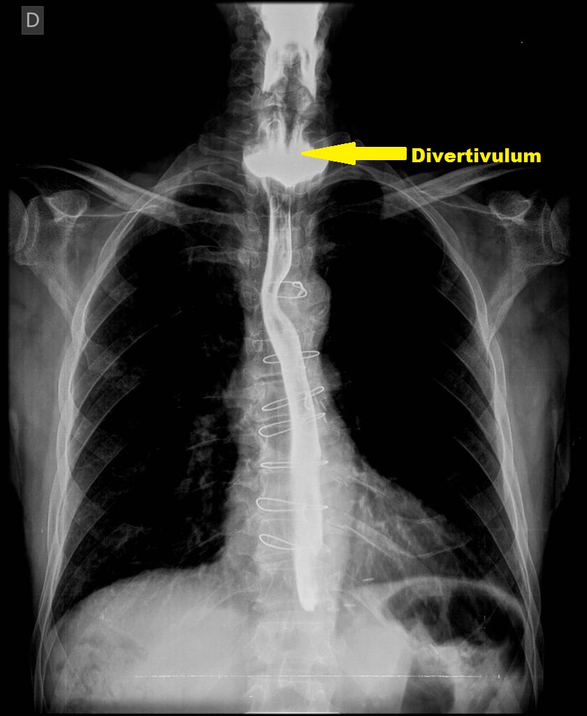 Source:Radiopaedia[1] |
|
| ||
| Esophageal carcinoma |
|
+ | + | Progressive | + | +/- | Normal |
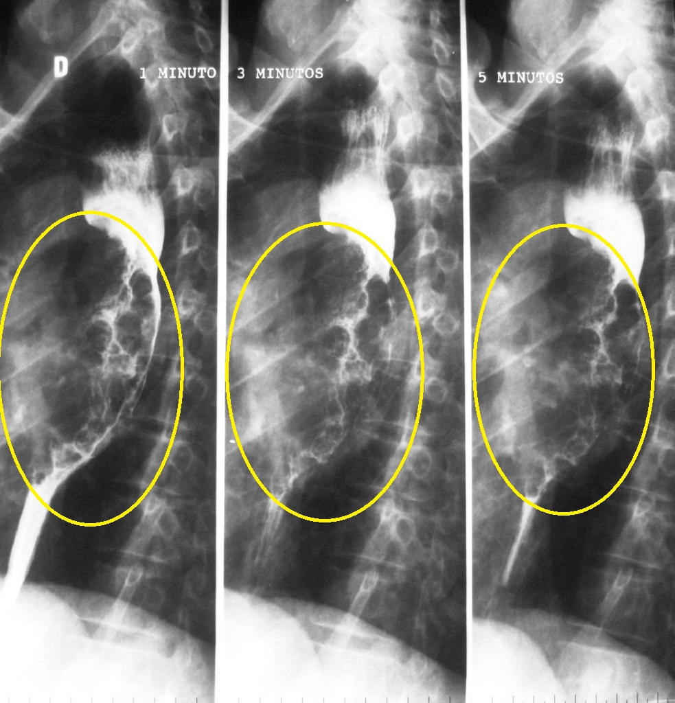 |
|
|||
| Stroke |
|
+ | + | Progressive | + | +/- |
|
Impaired |
 |
|
|
|
| Motor disorders |
|
+ | + | Progressive | +/- | Normal |
 |
|
|
| ||
| GERD |
|
+ | - | Progressive | +/- | + | Normal |
 |
|
| ||
| Esophageal web |
|
+ | +/- | Progressive | - | +/- |
|
Normal |
 |
|
|
|