Right bundle branch block electrocardiogram: Difference between revisions
Jump to navigation
Jump to search
No edit summary |
|||
| Line 21: | Line 21: | ||
== EKG Examples== | == EKG Examples== | ||
{| align="center" | |||
|-valign="top" | |||
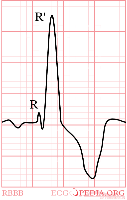
Image:RBBB1.png|The main characteristics of [[Right Bundle Branch Block]] in V1 | | [[Image:RBBB1.png|thumb|The main characteristics of [[Right Bundle Branch Block]] in V1]] | ||
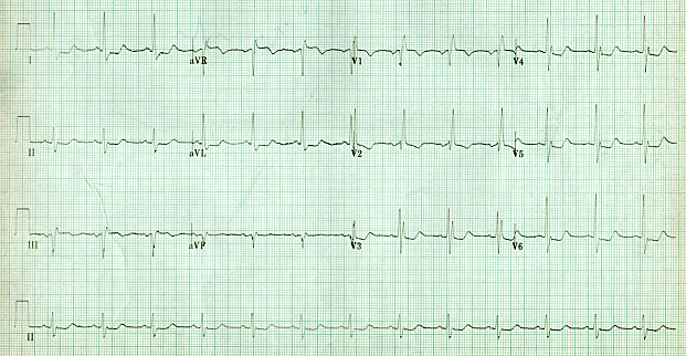
Image:ECG RBTB LAtrD.jpg|[[Right Bundle Branch Block]] | | [[Image:ECG RBTB LAtrD.jpg|thumb|[[Right Bundle Branch Block]]]] | ||
|} | |||
{| align="center" | |||
|-valign="top" | |||
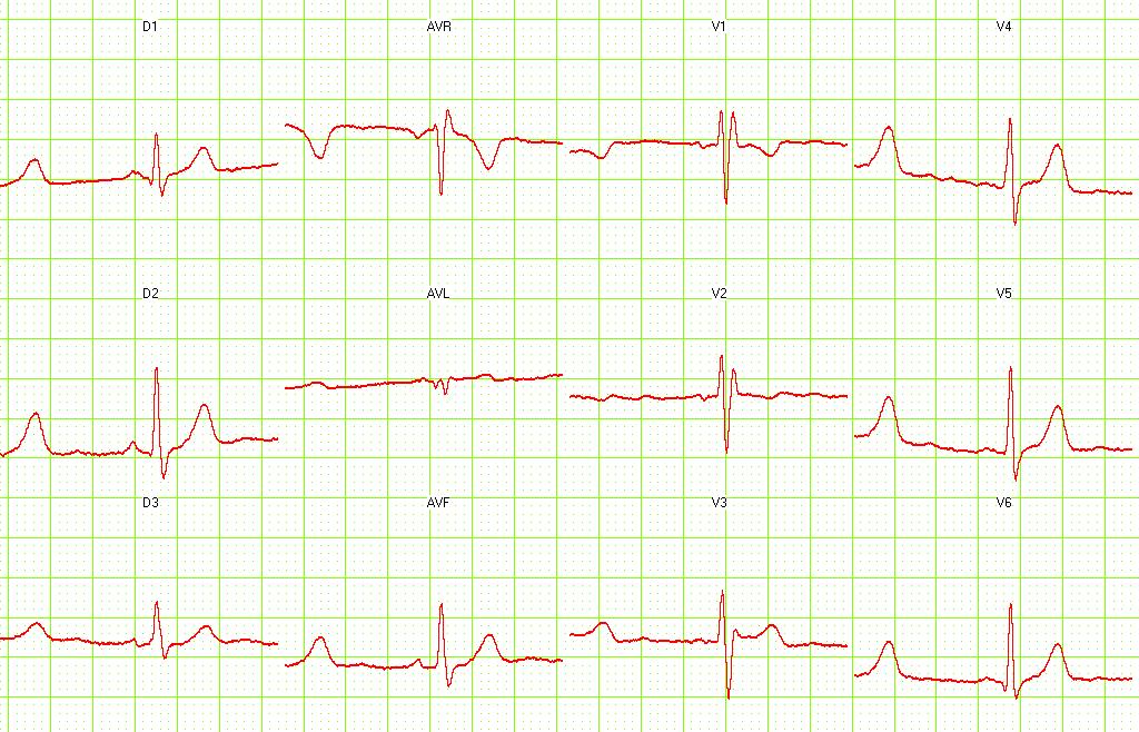
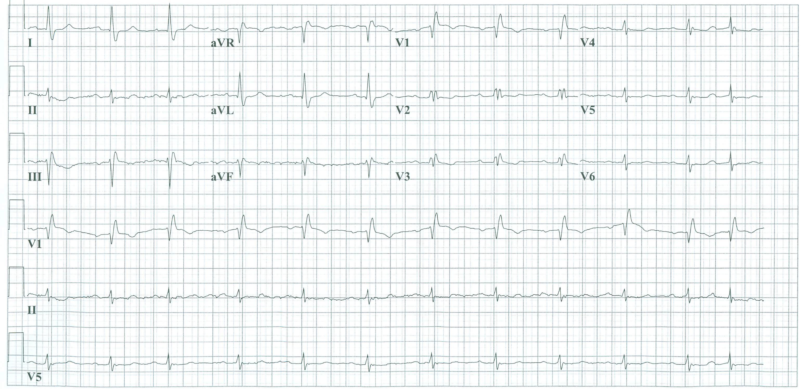
Image:RBBB.PNG|[[Right Bundle Branch Block]] | | [[Image:RBBB.PNG|thumb|[[Right Bundle Branch Block]]]] | ||
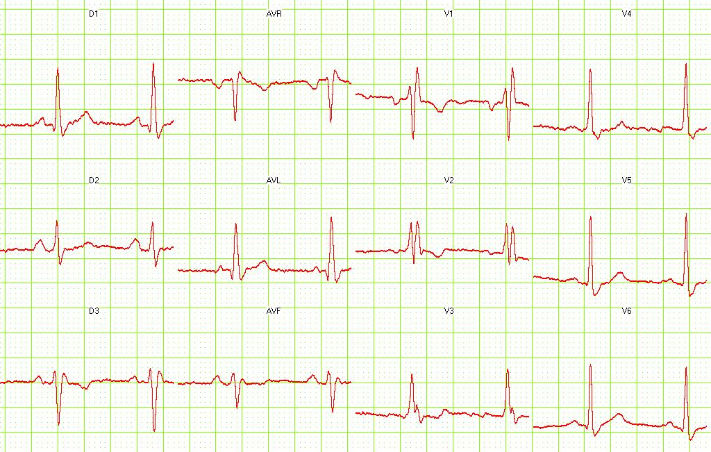
Image:C13.ht13.jpg|[[Right Bundle Branch Block]] | | [[Image:C13.ht13.jpg|thumb|[[Right Bundle Branch Block]]]] | ||
|} | |||
{| align="center" | |||
|-valign="top" | |||
Image:C14.ht14.jpg|[[Right Bundle Branch Block]]. <small> [http://www.ganseman.com/ecgbibnl.htm#_top000 Image courtesy of Dr Jose Ganseman]</small> | | [[Image:C14.ht14.jpg|thumb|[[Right Bundle Branch Block]]. <small> [http://www.ganseman.com/ecgbibnl.htm#_top000 Image courtesy of Dr Jose Ganseman]</small>]] | ||
Image:C15.ht15.jpg|[[Right Bundle Branch Block]]. <small> [http://www.ganseman.com/ecgbibnl.htm#_top000 Image courtesy of Dr Jose Ganseman]</small> | | [[Image:C15.ht15.jpg|thumb|[[Right Bundle Branch Block]]. <small> [http://www.ganseman.com/ecgbibnl.htm#_top000 Image courtesy of Dr Jose Ganseman]</small>]] | ||
|} | |||
{| align="center" | |||
|-valign="top" | |||
Image:C16.ht16.jpg|[[Right Bundle Branch Block]]. <small> [http://www.ganseman.com/ecgbibnl.htm#_top000 Image courtesy of Dr Jose Ganseman]</small> | | [[Image:C16.ht16.jpg|thumb|[[Right Bundle Branch Block]]. <small> [http://www.ganseman.com/ecgbibnl.htm#_top000 Image courtesy of Dr Jose Ganseman]</small>]] | ||
Image:C17.ht17.jpg|[[Right Bundle Branch Block]]. <small> [http://www.ganseman.com/ecgbibnl.htm#_top000 Image courtesy of Dr Jose Ganseman]</small> | | [[Image:C17.ht17.jpg|thumb|[[Right Bundle Branch Block]]. <small> [http://www.ganseman.com/ecgbibnl.htm#_top000 Image courtesy of Dr Jose Ganseman]</small>]] | ||
|} | |||
{| align="center" | |||
|-valign="top" | |||
Image:C18.ht18.jpg|[[Right Bundle Branch Block]] with [[First Degree AV Block|first degree AV block]]. <small> [http://www.ganseman.com/ecgbibnl.htm#_top000 Image courtesy of Dr Jose Ganseman]</small> | | [[Image:C18.ht18.jpg|thumb|[[Right Bundle Branch Block]] with [[First Degree AV Block|first degree AV block]]. <small> [http://www.ganseman.com/ecgbibnl.htm#_top000 Image courtesy of Dr Jose Ganseman]</small>]] | ||
Image:C22.ht22.jpg|[[Right Bundle Branch Block]] with RA hypertrophy. <small> [http://www.ganseman.com/ecgbibnl.htm#_top000 Image courtesy of Dr Jose Ganseman]</small> | | [[Image:C22.ht22.jpg|thumb|[[Right Bundle Branch Block]] with RA hypertrophy. <small> [http://www.ganseman.com/ecgbibnl.htm#_top000 Image courtesy of Dr Jose Ganseman]</small>]] | ||
|} | |||
{| align="center" | |||
|-valign="top" | |||
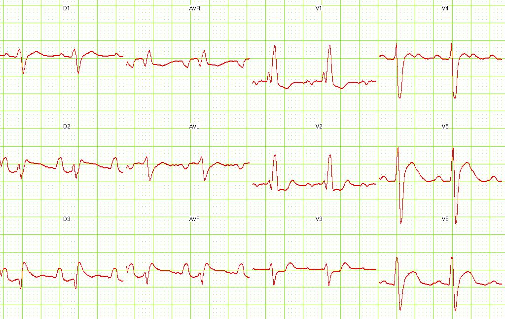
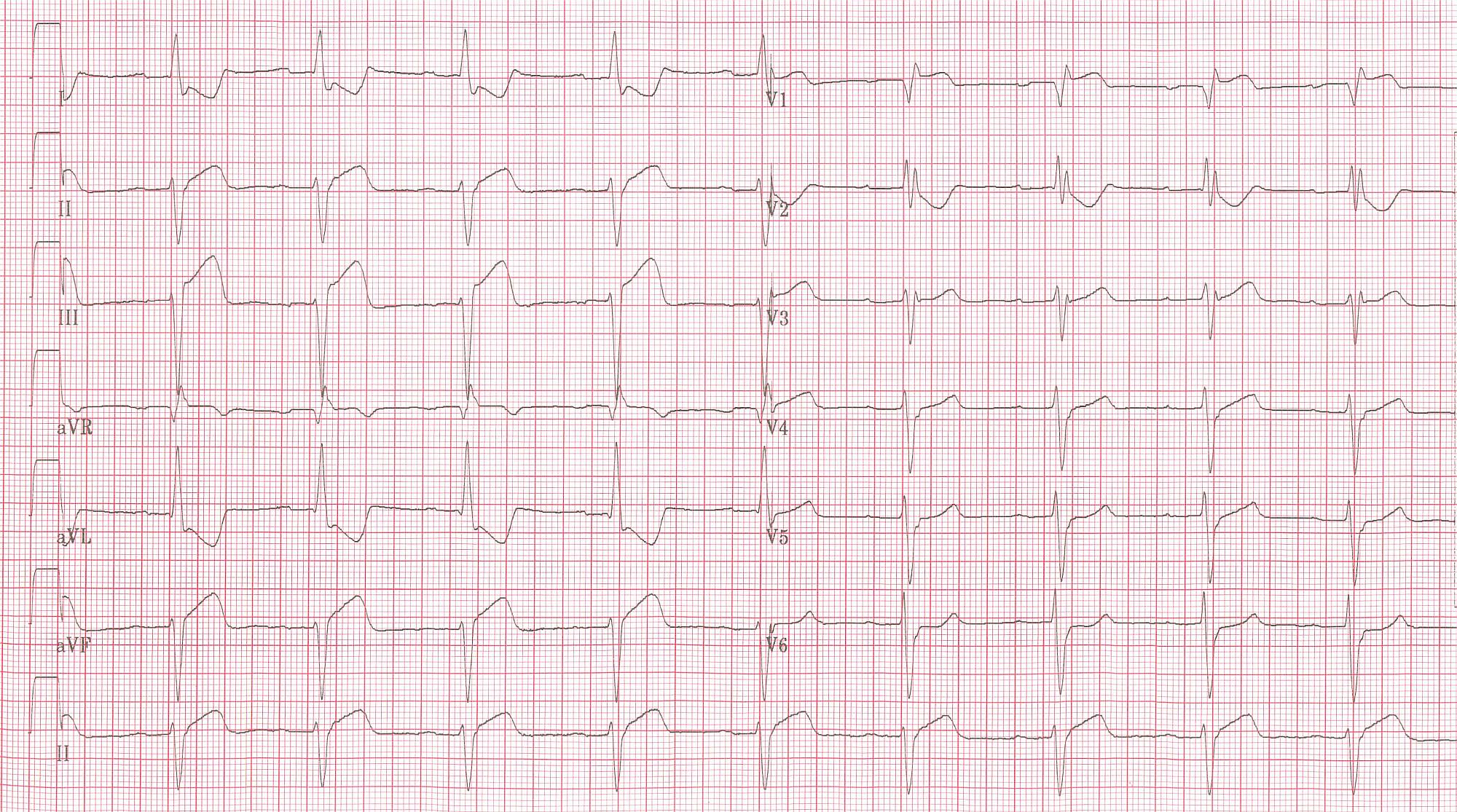
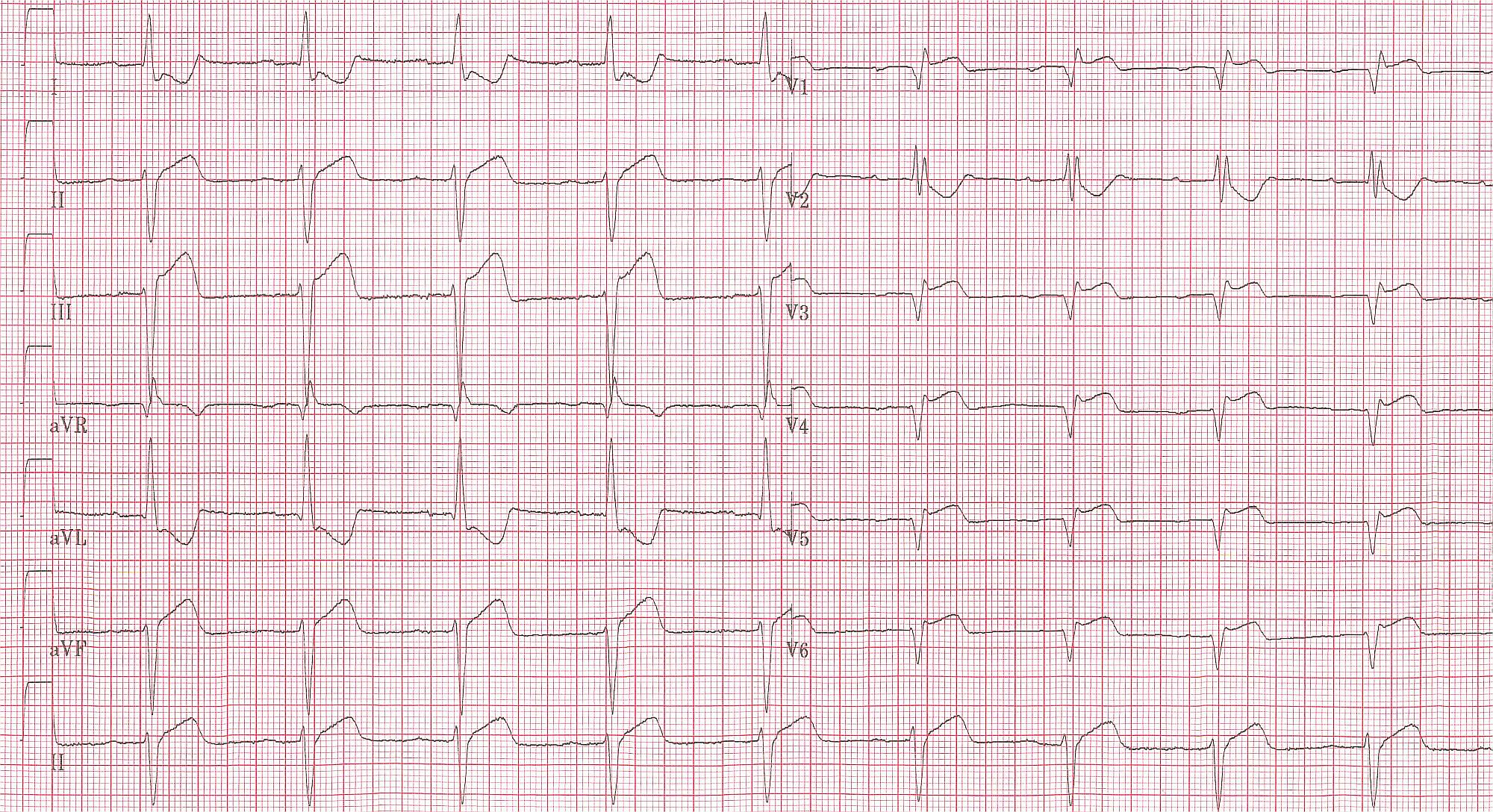
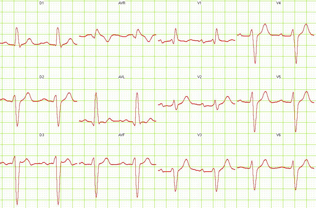
Image:RBBB_inf_MI.jpg|Patient with [[RBBB]] and [[Acute MI|inferior MI]]. Note to left axis deviation. | | [[Image:RBBB_inf_MI.jpg|thumb|Patient with [[RBBB]] and [[Acute MI|inferior MI]]. Note to left axis deviation.]] | ||
Image:RBBB_inf_MI_V4R.jpg|The same patient. Lead V4R. ST elevation shown. | | [[Image:RBBB_inf_MI_V4R.jpg|thumb|The same patient. Lead V4R. ST elevation shown.]] | ||
|} | |||
{| align="center" | |||
|-valign="top" | |||
Image:RBBB_inf_MI_baseline.jpg|The same patient before [[acute MI]] developed. Horizontal axis shown. | | [[Image:RBBB_inf_MI_baseline.jpg|thumb|The same patient before [[acute MI]] developed. Horizontal axis shown.]] | ||
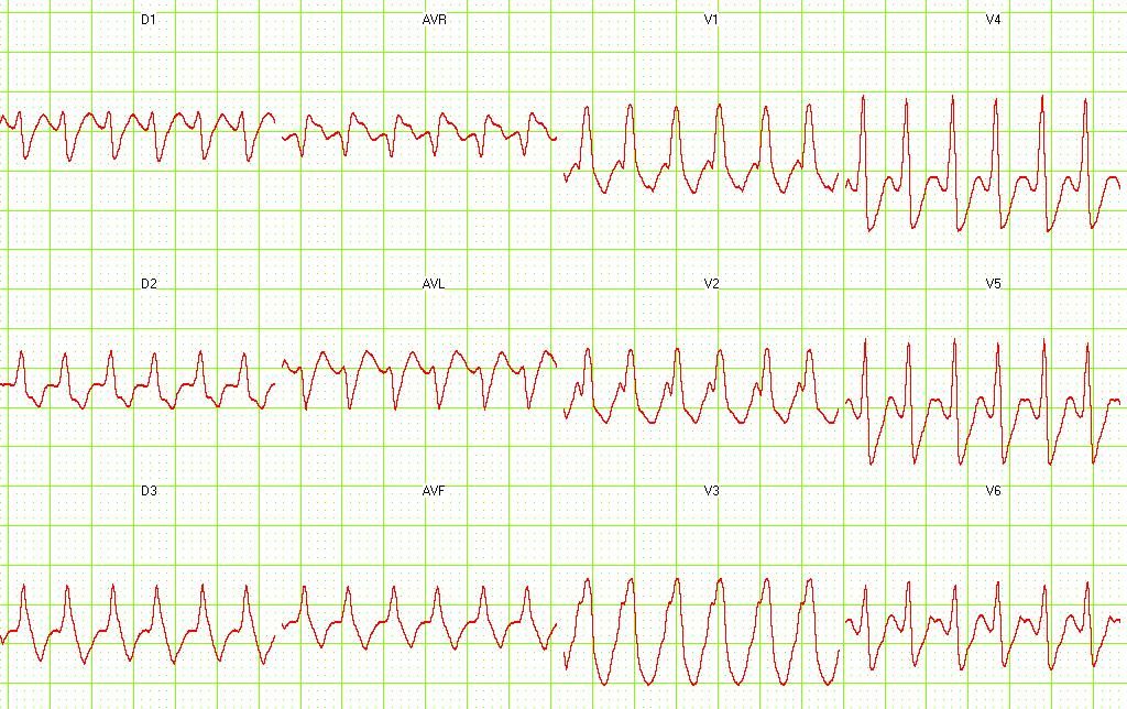
Image:R11.ht36.jpg|[[Supraventricular tachycardia]] with [[RBBB]]. <small> [http://www.ganseman.com/ecgbibnl.htm#_top000 Image courtesy of Dr Jose Ganseman]</small> | | [[Image:R11.ht36.jpg|thumb|[[Supraventricular tachycardia]] with [[RBBB]]. <small> [http://www.ganseman.com/ecgbibnl.htm#_top000 Image courtesy of Dr Jose Ganseman]</small>]] | ||
|} | |||
{| align="center" | |||
|-valign="top" | |||
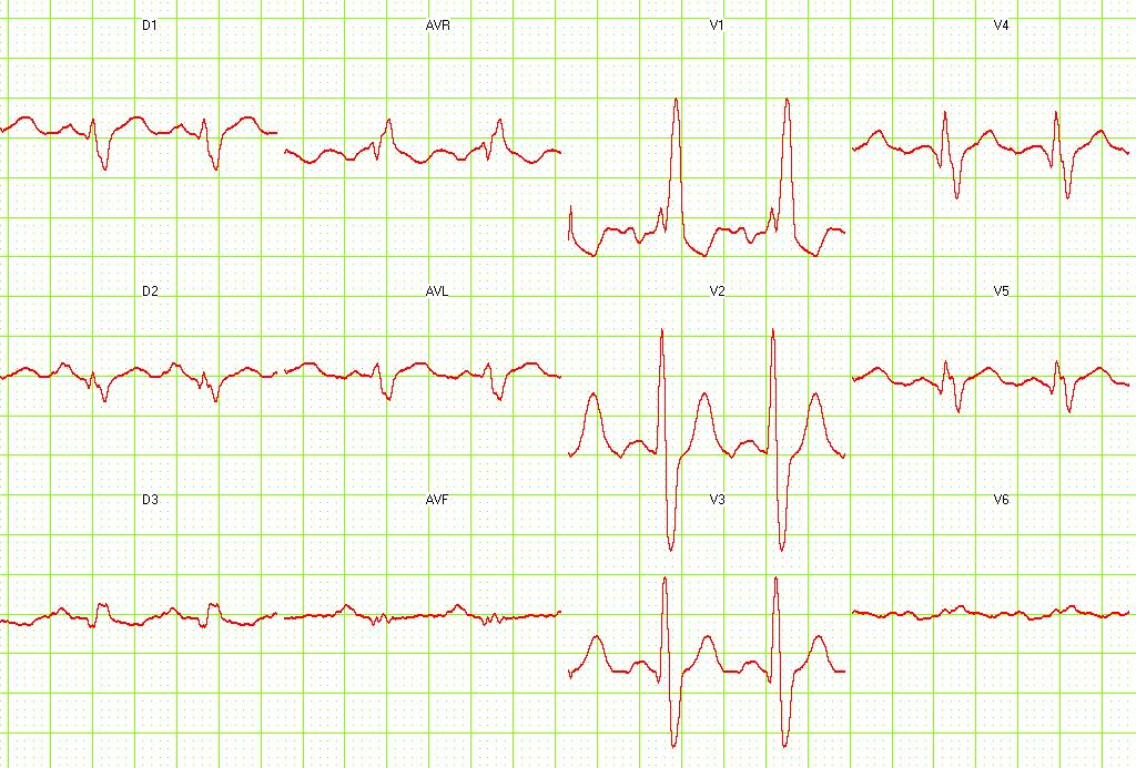
Image:cominf12.jpg|Old [[Acute MI|Anterior MI]] with [[RBBB]]. <small> [http://www.ganseman.com/ecgbibnl.htm#_top000 Image courtesy of Dr Jose Ganseman]</small> | | [[Image:cominf12.jpg|thumb|Old [[Acute MI|Anterior MI]] with [[RBBB]]. <small> [http://www.ganseman.com/ecgbibnl.htm#_top000 Image courtesy of Dr Jose Ganseman]</small>]] | ||
Image:cominf19.jpg|Old [[Acute MI|Inferior MI]] and [[Acute MI|Anterior MI] with [[RBBB]] and [[LAFB]]. | | [[Image:cominf19.jpg|thumb|Old [[Acute MI|Inferior MI]] and [[Acute MI|Anterior MI]] with [[RBBB]] and [[LAFB]].]] | ||
|} | |||
{| align="center" | |||
|-valign="top" | |||
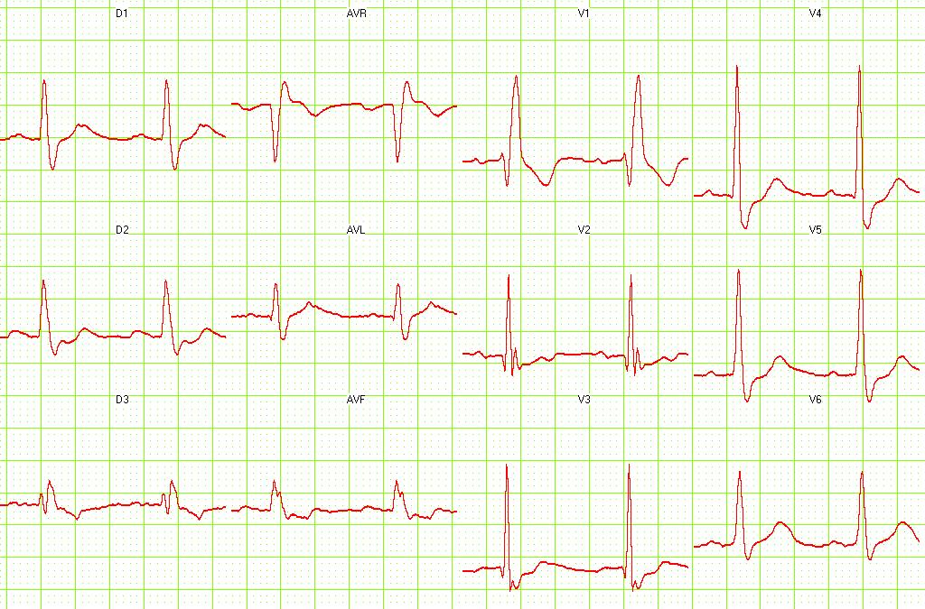
Image:cominf5.jpg|Old [[Acute MI|Inferior MI]] and [[RBBB]]. <small> [http://www.ganseman.com/ecgbibnl.htm#_top000 Image courtesy of Dr Jose Ganseman]</small> | | [[Image:cominf5.jpg|thumb|Old [[Acute MI|Inferior MI]] and [[RBBB]]. <small> [http://www.ganseman.com/ecgbibnl.htm#_top000 Image courtesy of Dr Jose Ganseman]</small>]] | ||
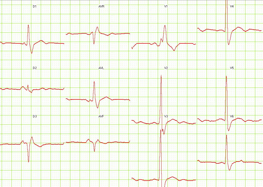
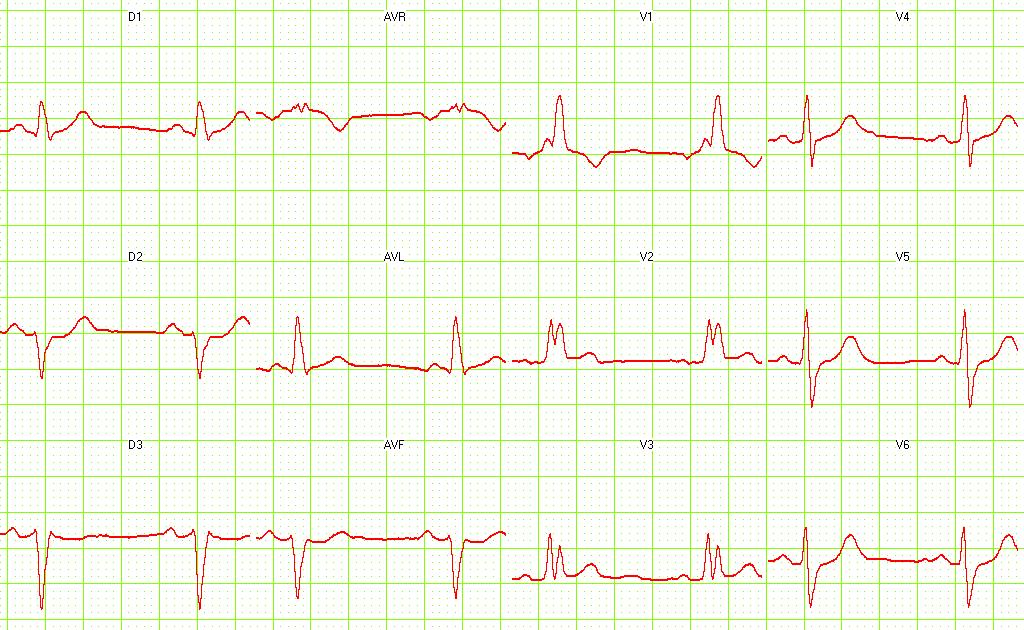
Image:c3.htm3.jpg|[[RBBB]] + [[LAFB]]. <small> [http://www.ganseman.com/ecgbibnl.htm#_top000 Image courtesy of Dr Jose Ganseman]</small> | | [[Image:c3.htm3.jpg|thumb|[[RBBB]] + [[LAFB]]. <small> [http://www.ganseman.com/ecgbibnl.htm#_top000 Image courtesy of Dr Jose Ganseman]</small>]] | ||
|} | |||
{| align="center" | |||
|-valign="top" | |||
Image:c19.ht19.jpg|[[RBBB]] + [[LAFB]] + [[First Degree AV Block]]. <small> [http://www.ganseman.com/ecgbibnl.htm#_top000 Image courtesy of Dr Jose Ganseman]</small> | | [[Image:c19.ht19.jpg|thumb|[[RBBB]] + [[LAFB]] + [[First Degree AV Block]]. <small> [http://www.ganseman.com/ecgbibnl.htm#_top000 Image courtesy of Dr Jose Ganseman]</small>]] | ||
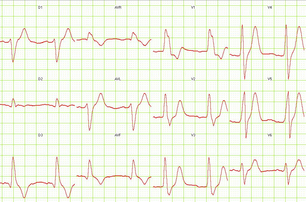
Image:c20.ht20.jpg|[[RBBB]] + [[LAFB]]. <small> [http://www.ganseman.com/ecgbibnl.htm#_top000 Image courtesy of Dr Jose Ganseman]</small> | | [[Image:c20.ht20.jpg|thumb|[[RBBB]] + [[LAFB]]. <small> [http://www.ganseman.com/ecgbibnl.htm#_top000 Image courtesy of Dr Jose Ganseman]</small>]] | ||
| [[Image:c21.ht21.jpg|thumb|[[RBBB]] + [[LPFB]]. <small> [http://www.ganseman.com/ecgbibnl.htm#_top000 Image courtesy of Dr Jose Ganseman]</small>]] | |||
|} | |||
Image:c21.ht21.jpg|[[RBBB]] + [[LPFB]]. <small> [http://www.ganseman.com/ecgbibnl.htm#_top000 Image courtesy of Dr Jose Ganseman]</small> | |||
==References== | ==References== | ||
Revision as of 15:04, 24 August 2012
|
Right bundle branch block Microchapters |
|
Differentiating Right bundle branch block from other Diseases |
|---|
|
Diagnosis |
|
Treatment |
|
Case Studies |
|
Right bundle branch block electrocardiogram On the Web |
|
American Roentgen Ray Society Images of Right bundle branch block electrocardiogram |
|
Risk calculators and risk factors for Right bundle branch block electrocardiogram |
Editor-In-Chief: C. Michael Gibson, M.S., M.D. [1] Associate Editor(s)-in-Chief: Cafer Zorkun, M.D., Ph.D. [2]
Overview
ECG
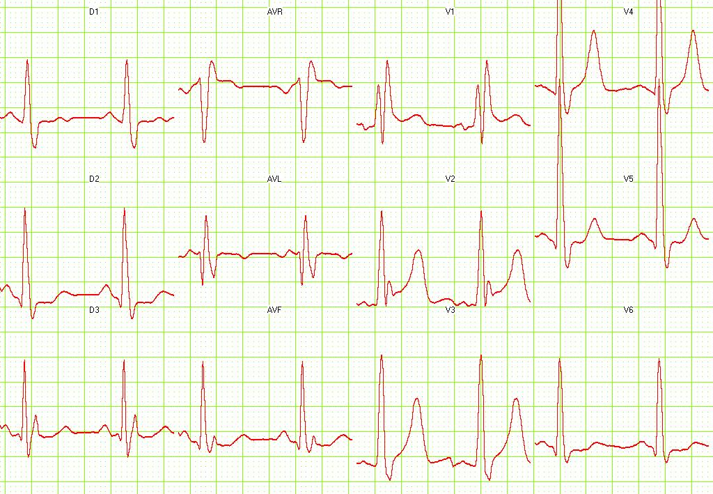
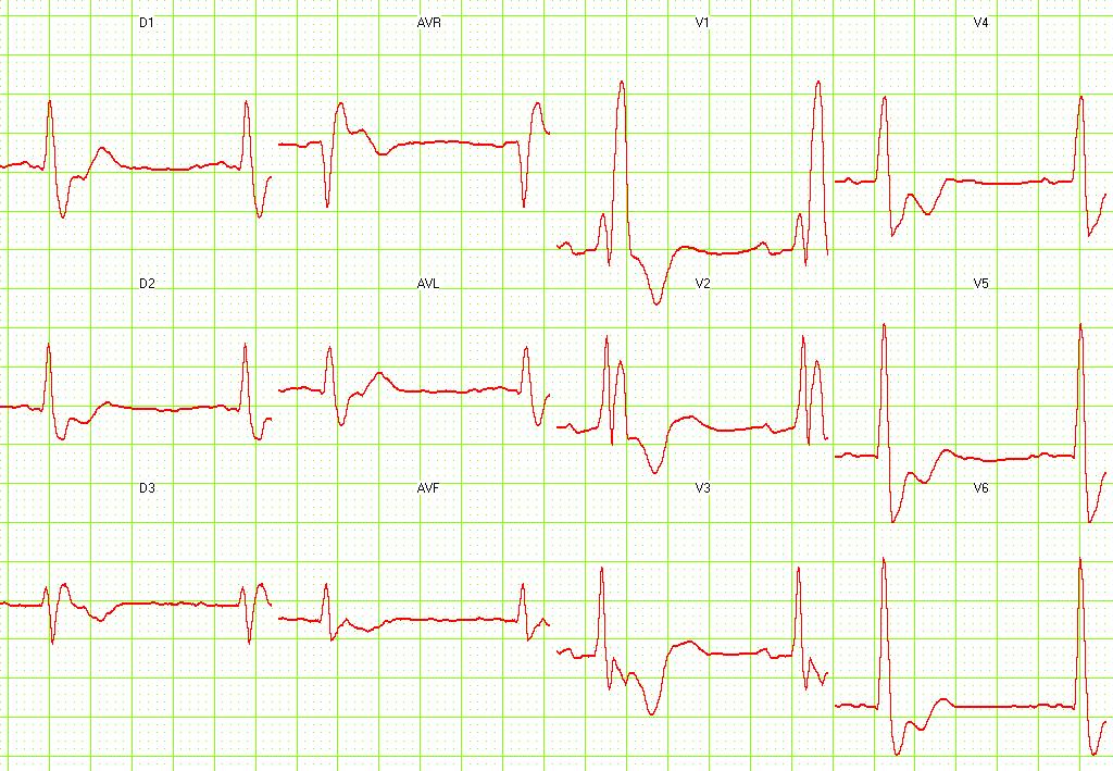
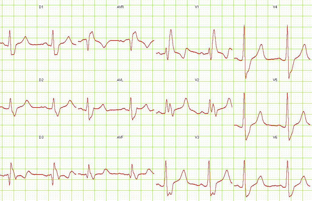
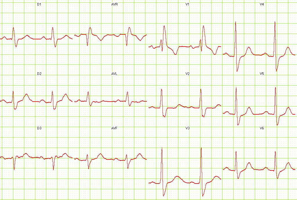
- The heart rhythm must be supraventricular in origin
- The QRS axis can be either normal, or right or left axis deviation may be present.
- The QRS duration must be = or > 120 ms
- For complete RBBB, the patient's age must be taken into account to determine if the duration of the QRS complex is prolonged for the patient's age.
- Maximum QRS durations are 0.07 s for newborns <6 days, 0.08 s for patients aged 1 week to 7 years, and 0.09 s for patients aged 7-15 years.
- For complete RBBB, the patient's age must be taken into account to determine if the duration of the QRS complex is prolonged for the patient's age.
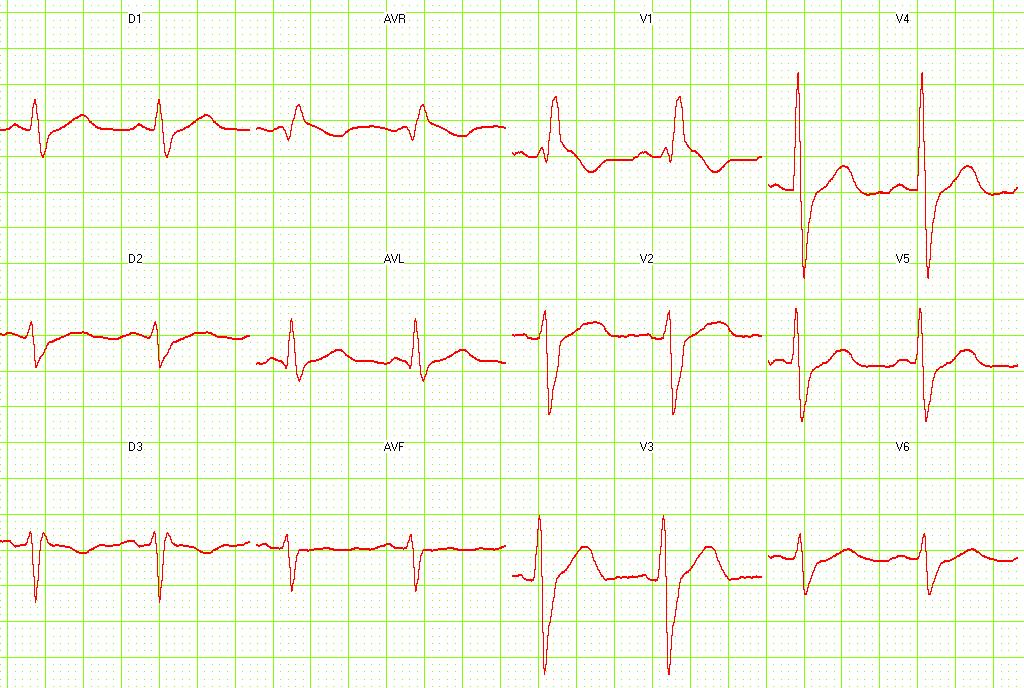
- There should be a terminal R wave in lead V1-V3R (e.g., R, rR', rsR', rSR' or qR')
- This pattern is present because the initial R wave represents septal activation, the S wave represents left ventricular activation, and the R' represents activation of the right ventricle from the septum and left ventricle.
- There should be a slurred S wave in leads I and V6. This represent left ventricular activation.
- Because transmission of the electrical impulse through the left bundle is normal, this results in normal depolarization of the septum and the left ventricle. As a result, there is an initial R wave in lead I and V1 and the Q wave in V6.
The T wave should be deflected opposite the terminal deflection of the QRS complex. This is known as appropriate T wave discordance with bundle branch block. A concordant T wave may suggest ischemia or myocardial infarction.
EKG Examples
 |
 |
 |
 |
 |
 |
 |
 |
 |
 |
 |
 |
 |
 |
 |
 |
 |
 |
 |
 |