Psoriasis physical examination: Difference between revisions
Jump to navigation
Jump to search
No edit summary |
No edit summary |
||
| Line 27: | Line 27: | ||
[[Image:Psoriahair.jpg|200px|align right|scalp psoriasis|courtesy regionalderm.net]] | [[Image:Psoriahair.jpg|200px|align right|scalp psoriasis|courtesy regionalderm.net]] | ||
====Extremities==== | ====Extremities==== | ||
[[Image:Extremity_psoriasis.jpg|200px|align right|extremity psoriasis(guttate variety)|courtesy regionalderm.net]] | |||
=====Trunk===== | =====Trunk===== | ||
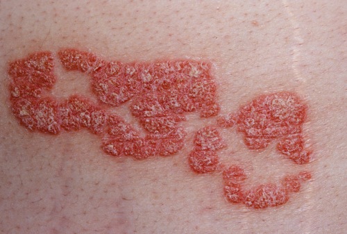
[[Image:Trunkpsor.jpg|200px|align right|trunk psoriasis|courtesy regionalderm.net]] | [[Image:Trunkpsor.jpg|200px|align right|trunk psoriasis|courtesy regionalderm.net]] | ||
=====Face===== | =====Face===== | ||
[[Image:Face. | [[Image:Face.jpg|200px|align right|face psoriasis]] | ||
=====Nail Psoriasis===== | =====Nail Psoriasis===== | ||
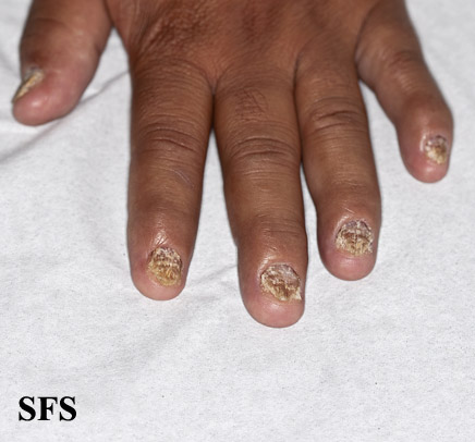
[[Image:Nail_psoriasis01.jpg|200px|align right|Nails showing pitting, crumbling and brittleness]] | [[Image:Nail_psoriasis01.jpg|200px|align right|Nails showing pitting, crumbling and brittleness]] | ||
=====Inverse Psoriasis===== | =====Inverse Psoriasis===== | ||
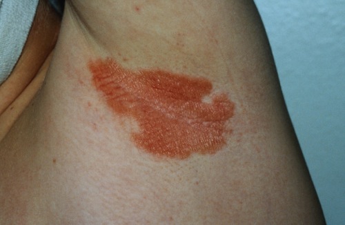
[[Image:PsoriasisAxilla.jpg|200px|align right|inverse psoriasis|courtesy regionalderm.net]] | [[Image:PsoriasisAxilla.jpg|200px|align right|inverse psoriasis|courtesy regionalderm.net]] | ||
===HEENT=== | ===HEENT=== | ||
*Scalp psoriasis may cause raised, reddish, often scaly patches. | *Scalp psoriasis may cause raised, reddish, often scaly patches. | ||
Revision as of 15:54, 5 July 2017
|
Psoriasis Microchapters |
|
Diagnosis |
|---|
|
Treatment |
|
Case Studies |
|
Psoriasis physical examination On the Web |
|
American Roentgen Ray Society Images of Psoriasis physical examination |
|
Risk calculators and risk factors for Psoriasis physical examination |
Editor-In-Chief: C. Michael Gibson, M.S., M.D. [1]; Associate Editor(s)-in-Chief: Syed Hassan A. Kazmi BSc, MD [2] Kiran Singh, M.D. [3]
Overview
Common physical examination findings of psoraisis include erythematous, scaling papules and plaques on the skin.
Physical Examination
Appearance of the Patient
- Patient with psoriasis may look distressed and anxious
Vital signs
- High-grade fever with generalized pustular psoriasis.
- Tachycardia with regular pulse.
- Tachypnea.
- Kussmal respirations may be present in patients with comorbid diabetes and DKA.
- High-output cardiac failure in erythroderma.
Skin
- A diagnosis of psoriasis is usually based on the appearance of the skin. There are no special blood tests or diagnostic procedures for psoriasis. Sometimes a skin biopsy, or scraping, may be needed to rule out other disorders and to confirm the diagnosis. Skin from a biopsy will show clubbed rete pegs if positive for psoriasis.
- Psoriasis is a papulosquamous disease with variable morphology, distribution, severity, and course.
- It is characterized by scaling papules and plaques.
Scalp
Extremities
Trunk
Face
Nail Psoriasis
Inverse Psoriasis
HEENT
- Scalp psoriasis may cause raised, reddish, often scaly patches.
- Ophthalmoscopic exam in psoriasis may show uveitis, more frequently in patients with arthropathy or pustular psoriasis.[1]
- Sensorineural hearing loss associated with psoriatic arthritis.
- Rinne test may be negative (abnormal).
- Weber test may show a quieter sound in the ear with the sensorineuronal hearing loss.
Neck
- Cervical Lymphadenopathy
Lungs
- Psoriasis has been known to be associated with COPD.[2]
- Exapnded/barrel shaped chest because of COPD.
- Bilateral decresed breath sounds.
- Bilateral wheezes.
- Egophony absent.
- Reduced tactile fremitus.
Heart
- The risk of arterial and venous vascular diseases (eg, myocardial infarction, thrombophlebitis, pulmonary embolization) is higher in sever psoriasis involving multiple areas of the body.[3]
- There may be a chance of getting high output cardiac failure to to erytheroderma.[3]
===Abdomen===.
- No abdominal distention.
- No abdominal tenderness.
- No Hepatomegaly / splenomegaly / hepatosplenomegaly.
References
- ↑ Fraga NA, Oliveira Mde F, Follador I, Rocha Bde O, Rêgo VR (2012). "Psoriasis and uveitis: a literature review". An Bras Dermatol. 87 (6): 877–83. PMC 3699904. PMID 23197207.
- ↑ Dreiher J, Weitzman D, Shapiro J, Davidovici B, Cohen AD (2008). "Psoriasis and chronic obstructive pulmonary disease: a case-control study". Br. J. Dermatol. 159 (4): 956–60. doi:10.1111/j.1365-2133.2008.08749.x. PMID 18637897.
- ↑ 3.0 3.1 Kremers HM, McEvoy MT, Dann FJ, Gabriel SE (2007). "Heart disease in psoriasis". J. Am. Acad. Dermatol. 57 (2): 347–54. doi:10.1016/j.jaad.2007.02.007. PMID 17433490.


