Plummer-Vinson syndrome x ray: Difference between revisions
Akshun Kalia (talk | contribs) (→X Ray) |
Akshun Kalia (talk | contribs) No edit summary |
||
| Line 1: | Line 1: | ||
__NOTOC__ | __NOTOC__ | ||
{{Plummer-Vinson syndrome}} | {{Plummer-Vinson syndrome}} | ||
{{CMG}} {{AE}} | {{CMG}};{{AE}}{{Akshun}} | ||
{{ | |||
==Overview== | ==Overview== | ||
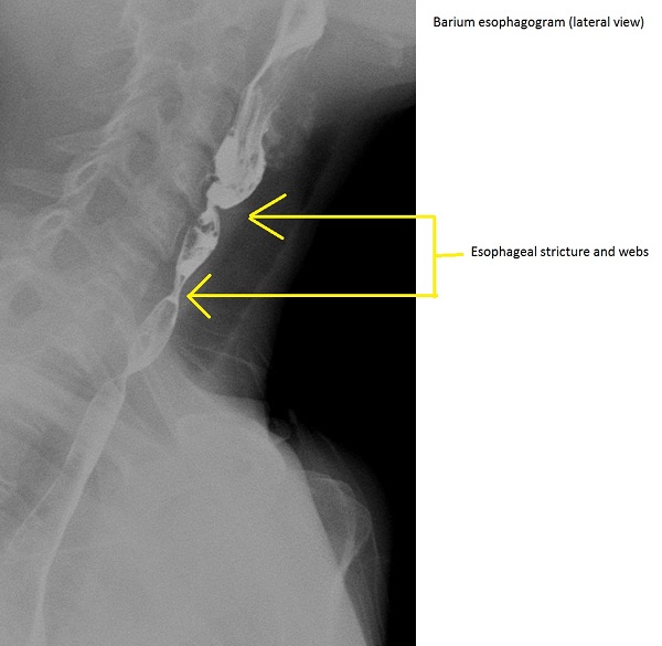
An x-ray (barium esophagogram) is the best initial imaging study in a patient suspected with Plummer-Vinson syndrome. Findings on an x-ray (barium esophagogram) suggestive of esophageal web/strictures associated with Plummer-Vinson syndrome appear as either thin projections on the anterior esophageal wall or multiple upper (cervical) esophageal constrictions consistent with esophageal webs. | |||
==X Ray== | ==X Ray== | ||
Revision as of 13:55, 30 October 2017
|
Plummer-Vinson syndrome Microchapters |
|
Diagnosis |
|---|
|
Treatment |
|
Case Studies |
|
Plummer-Vinson syndrome x ray On the Web |
|
American Roentgen Ray Society Images of Plummer-Vinson syndrome x ray |
|
Risk calculators and risk factors for Plummer-Vinson syndrome x ray |
Editor-In-Chief: C. Michael Gibson, M.S., M.D. [1];Associate Editor(s)-in-Chief: Akshun Kalia M.B.B.S.[2]
Overview
An x-ray (barium esophagogram) is the best initial imaging study in a patient suspected with Plummer-Vinson syndrome. Findings on an x-ray (barium esophagogram) suggestive of esophageal web/strictures associated with Plummer-Vinson syndrome appear as either thin projections on the anterior esophageal wall or multiple upper (cervical) esophageal constrictions consistent with esophageal webs.
X Ray
- An x-ray is the best initial test and can be helpful in the diagnosis of Plummer-Vinson syndrome.
- A barium esophagogram helps in determining the calibre of esophageal lumen.
- Findings on an x-ray (barium esophagogram) suggestive of esophageal web/strictures associated with Plummer-Vinson syndrome appear as either:
- Thin projections on the anterior esophageal wall.
- Multiple upper (cervical) esophageal constrictions consistent with esophageal webs.
