Pleural effusion chest x ray: Difference between revisions
Ahmed Younes (talk | contribs) |
Ahmed Younes (talk | contribs) |
||
| Line 10: | Line 10: | ||
Image:Pleural effusion.jpg|'''Pleural effusion''' Chest x-ray of a pleural effusion. The arrow A shows fluid layering in the right pleural cavity. The B arrow shows the normal width of the lung in the cavity - Case courtesy of Dr Vivek Pai, <a href="https://radiopaedia.org/">Radiopaedia.org</a>. From the case <a href="https://radiopaedia.org/cases/27112">rID: 27112</a> | Image:Pleural effusion.jpg|'''Pleural effusion''' Chest x-ray of a pleural effusion. The arrow A shows fluid layering in the right pleural cavity. The B arrow shows the normal width of the lung in the cavity - Case courtesy of Dr Vivek Pai, <a href="https://radiopaedia.org/">Radiopaedia.org</a>. From the case <a href="https://radiopaedia.org/cases/27112">rID: 27112</a> | ||
Image:Right side unilateral effusion 1.jpg|Large unilateral right sided effusion. Heart is enlarged, especially the left lateral appendage. - Case courtesy of A.Prof Frank Gaillard, <a href="https://radiopaedia.org/">Radiopaedia.org</a>. From the case <a href="https://radiopaedia.org/cases/24290">rID: 24290</a> | Image:Right side unilateral effusion 1.jpg|Large unilateral right sided effusion. Heart is enlarged, especially the left lateral appendage. - Case courtesy of A.Prof Frank Gaillard, <a href="https://radiopaedia.org/">Radiopaedia.org</a>. From the case <a href="https://radiopaedia.org/cases/24290">rID: 24290</a> | ||
Image:Right side pleural effusion 2.png|Right side pleural effusion. A homogenous opacification is noted in the right lower zone. The right costophrenic angle is obliterated with a meniscus noted. - | Image:Right side pleural effusion 2.png|Right side pleural effusion. A homogenous opacification is noted in the right lower zone. The right costophrenic angle is obliterated with a meniscus noted. - Source: https://www.cdc.gov/ | ||
</gallery> | </gallery> | ||
'''Small bilateral pleural effusions that layer with decubitus views''' | '''Small bilateral pleural effusions that layer with decubitus views''' | ||
Revision as of 20:22, 21 September 2017
|
Pleural effusion Microchapters |
|
Diagnosis |
|---|
|
Treatment |
|
Case Studies |
|
Pleural effusion chest x ray On the Web |
|
American Roentgen Ray Society Images of Pleural effusion chest x ray |
|
Risk calculators and risk factors for Pleural effusion chest x ray |
Editor-In-Chief: C. Michael Gibson, M.S., M.D. [1] Associate Editor(s)-in-Chief: Prince Tano Djan, BSc, MBChB [2]
Overview
Chest films acquired in the lateral decubitus position (with the patient lying on their side) are more sensitive, and can detect as little as 50 ml of fluid. At least 200ml-300 ml of fluid must be present before upright chest films can detect signs of pleural effusion (e.g. blunted costophrenic angles).[1]
Chest X Ray
-
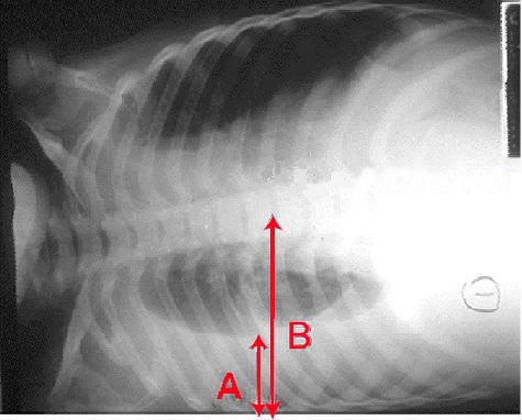
Pleural effusion Chest x-ray of a pleural effusion. The arrow A shows fluid layering in the right pleural cavity. The B arrow shows the normal width of the lung in the cavity - Case courtesy of Dr Vivek Pai, <a href="https://radiopaedia.org/">Radiopaedia.org</a>. From the case <a href="https://radiopaedia.org/cases/27112">rID: 27112</a>
-
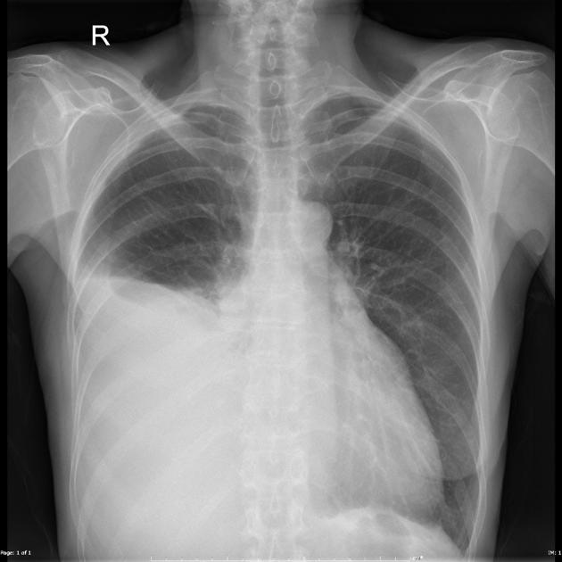
Large unilateral right sided effusion. Heart is enlarged, especially the left lateral appendage. - Case courtesy of A.Prof Frank Gaillard, <a href="https://radiopaedia.org/">Radiopaedia.org</a>. From the case <a href="https://radiopaedia.org/cases/24290">rID: 24290</a>
-
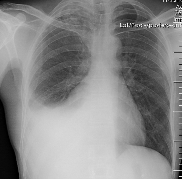
Right side pleural effusion. A homogenous opacification is noted in the right lower zone. The right costophrenic angle is obliterated with a meniscus noted. - Source: https://www.cdc.gov/


Small bilateral pleural effusions that layer with decubitus views


