Osteomyelitis MRI
|
Osteomyelitis Microchapters |
|
Diagnosis |
|---|
|
Treatment |
|
Case Studies |
|
Osteomyelitis MRI On the Web |
|
American Roentgen Ray Society Images of Osteomyelitis MRI |
Editor-In-Chief: C. Michael Gibson, M.S., M.D. [1]
Please help WikiDoc by adding more content here. It's easy! Click here to learn about editing.
Overview
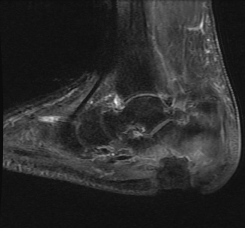
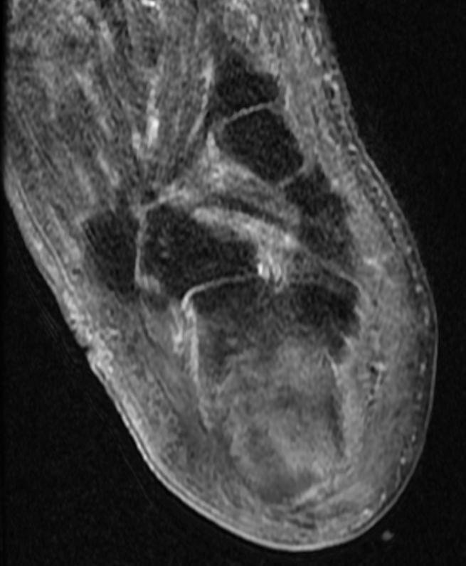
MR imaging is the accepted modality of choice for the early detection and surgical localization of osteomyelitis.
MRI
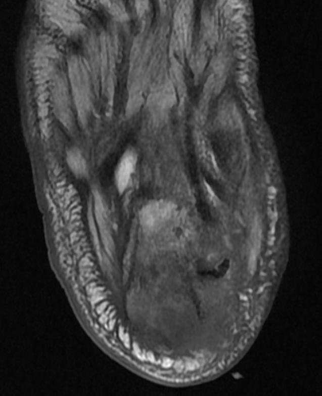
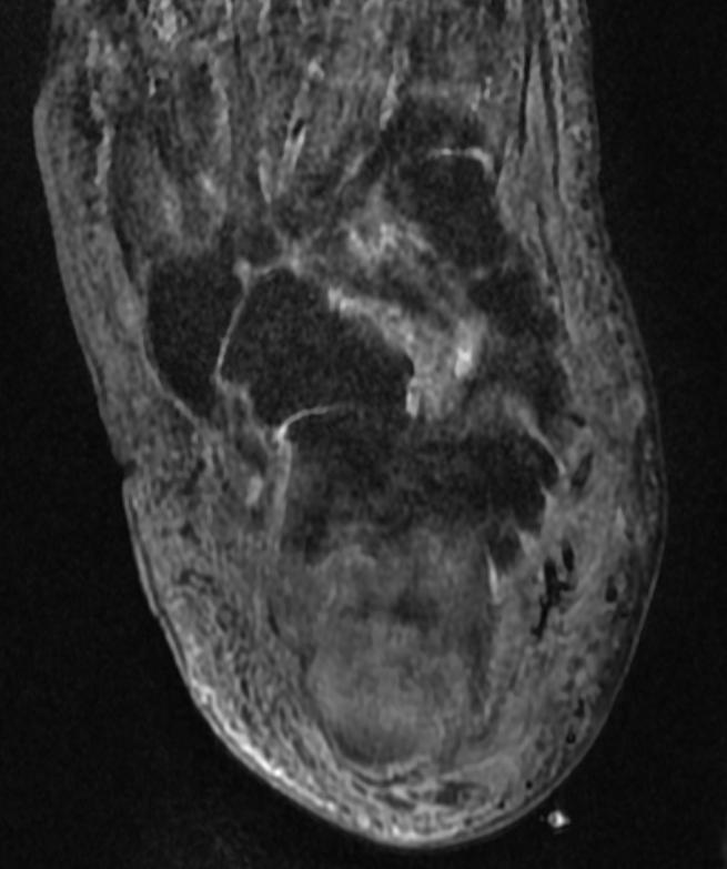
Patient #1 Extensive calcaneal osteomyelitis. Note soft tissue ulceration and cellulitis
-
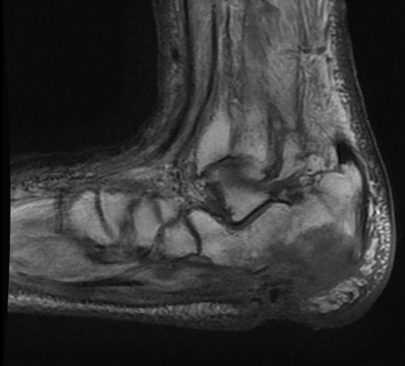
T1
-
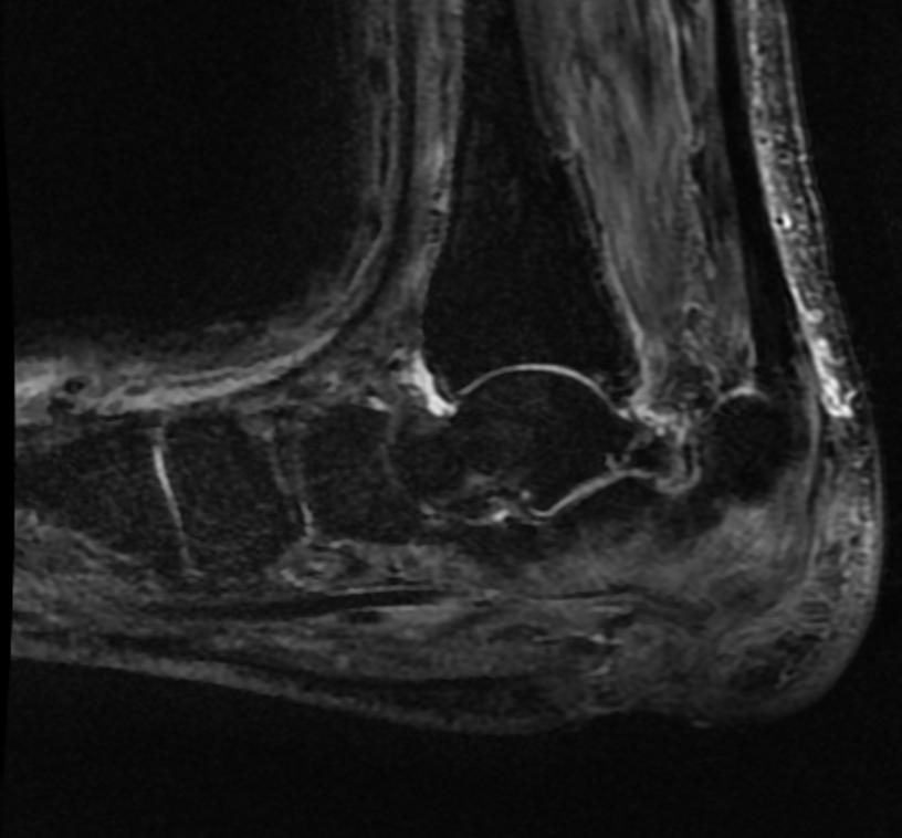
STIR
-
T1
-
STIR
-
T1 fat sat contrast
-
T1 fat sat contrast


