Multiple myeloma MRI
|
Multiple myeloma Microchapters |
|
Diagnosis |
|---|
|
Treatment |
|
Case Studies |
|
Multiple myeloma MRI On the Web |
|
American Roentgen Ray Society Images of Multiple myeloma MRI |
Editor-In-Chief: C. Michael Gibson, M.S., M.D. [1]
Overview
Magnetic resonance imaging (MRI) is more sensitive than simple X-ray in the detection of lytic lesions of multiple myeloma. MRI may supersede skeletal survey, especially when vertebral disease is suspected.
MRI
Shown below is a series of MRI images of long bones involved in multiple myeloma. (Images courtesy of RadsWiki)
-
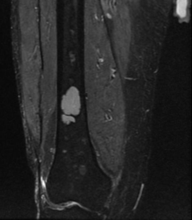
Multiple myeloma
-
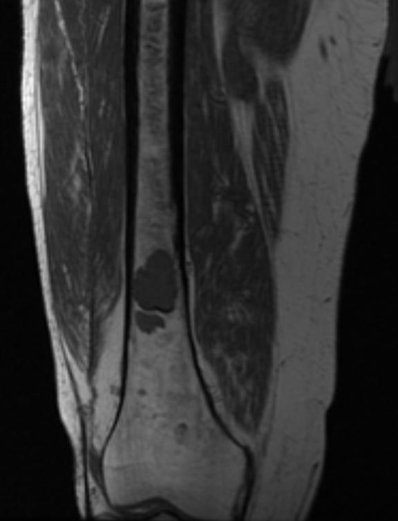
Multiple myeloma
-
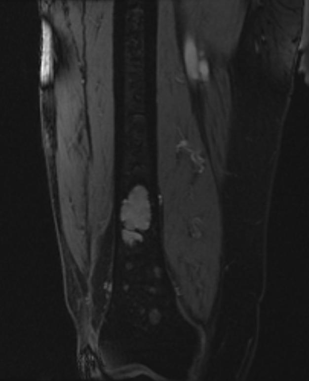
Multiple myeloma


Shown below is a series of MRI images in a multiple myeloma patient complaining of back pain.

