Multiple myeloma MRI: Difference between revisions
Varun Kumar (talk | contribs) No edit summary |
(→MRI) |
||
| Line 10: | Line 10: | ||
Image:Multiple_myeloma_MRI102.jpg|Multiple myeloma | Image:Multiple_myeloma_MRI102.jpg|Multiple myeloma | ||
Image:Multiple_myeloma_MRI103.jpg|Multiple myeloma | Image:Multiple_myeloma_MRI103.jpg|Multiple myeloma | ||
</gallery> | |||
'''Back pain in a patient with known multiple myeloma''' | |||
<gallery> | |||
Image: | |||
Multiple-myeloma-101.jpg | |||
Image: | |||
Multiple-myeloma-102.jpg | |||
</gallery> | </gallery> | ||
Revision as of 15:25, 5 August 2012
|
Multiple myeloma Microchapters |
|
Diagnosis |
|---|
|
Treatment |
|
Case Studies |
|
Multiple myeloma MRI On the Web |
|
American Roentgen Ray Society Images of Multiple myeloma MRI |
Editor-In-Chief: C. Michael Gibson, M.S., M.D. [1]
MRI
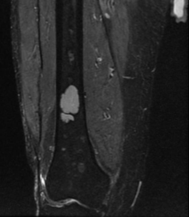
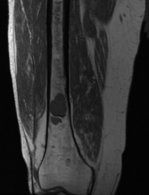
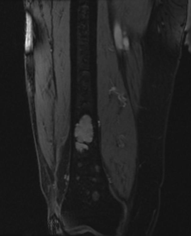
The workup of suspected multiple myeloma includes a skeletal survey. This is a series of X-rays of the skull, axial skeleton and proximal long bones. Myeloma activity sometimes appear as "lytic lesions" (with local disappearance of normal bone due to resorption), and on the skull X-ray as "punched-out lesions" (pepper pot skull). Magnetic resonance imaging (MRI) is more sensitive than simple X-ray in the detection of lytic lesions, and may supersede skeletal survey, especially when vertebral disease is suspected. Occasionally a CT scan is performed to measure the size of soft tissue plasmacytomas.
-
Multiple myeloma
-
Multiple myeloma
-
Multiple myeloma


Back pain in a patient with known multiple myeloma

