Medial palpebral arteries
| Cardiology Network |
 Discuss Medial palpebral arteries further in the WikiDoc Cardiology Network |
| Adult Congenital |
|---|
| Biomarkers |
| Cardiac Rehabilitation |
| Congestive Heart Failure |
| CT Angiography |
| Echocardiography |
| Electrophysiology |
| Cardiology General |
| Genetics |
| Health Economics |
| Hypertension |
| Interventional Cardiology |
| MRI |
| Nuclear Cardiology |
| Peripheral Arterial Disease |
| Prevention |
| Public Policy |
| Pulmonary Embolism |
| Stable Angina |
| Valvular Heart Disease |
| Vascular Medicine |
Editor-In-Chief: C. Michael Gibson, M.S., M.D. [1]
Overview
The medial palpebral arteries (internal palpebral arteries), two in number, superior and inferior, arise from the ophthalmic, opposite the pulley of the Obliquus superior; they leave the orbit to encircle the eyelids near their free margins, forming a superior and an inferior arch, which lie between the Orbicularis oculi and the tarsi.
The superior palpebral anastomoses, at the lateral angle of the orbit, with the zygomaticoörbital branch of the temporal artery and with the upper of the two lateral palpebral branches from the lacrimal artery.
The inferior palpebral anastomoses, at the lateral angle of the orbit, with the lower of the two lateral palpebral branches from the lacrimal and with the transverse facial artery, and, at the medial part of the lid, with a branch from the angular artery.
From this last anastomoses a branch passes to the nasolacrimal duct, ramifying in its mucous membrane, as far as the inferior meatus of the nasal cavity.
Additional images
-
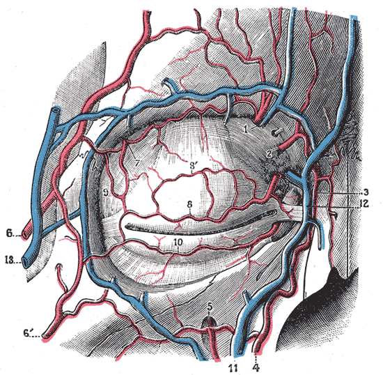
Bloodvessels of the eyelids, front view.
