Lymphadenopathy: Difference between revisions
No edit summary |
|||
| Line 202: | Line 202: | ||
'''Ultrasonography''' | '''Ultrasonography''' | ||
*On ultrasound, characteristic findings of lymphadenopathy, include:<ref name="pmid24753638">{{cite journal |vauthors=Mohseni S, Shojaiefard A, Khorgami Z, Alinejad S, Ghorbani A, Ghafouri A |title=Peripheral lymphadenopathy: approach and diagnostic tools |journal=Iran J Med Sci |volume=39 |issue=2 Suppl |pages=158–70 |year=2014 |pmid=24753638 |pmc=3993046 |doi= |url=}}</ref><ref name="radio">Lymph node enlargment. Radiopedia. http://radiopaedia.org/articles/lymph-node-enlargement Accessed on May 9, 2016 </ref><ref name="AA">Lymph node enlargment. Wikipedia. https://en.wikipedia.org/wiki/Lymph_node Accessed on May 9, 2016</ref> | *On ultrasound, characteristic findings of lymphadenopathy, include:<ref name="pmid24753638">{{cite journal |vauthors=Mohseni S, Shojaiefard A, Khorgami Z, Alinejad S, Ghorbani A, Ghafouri A |title=Peripheral lymphadenopathy: approach and diagnostic tools |journal=Iran J Med Sci |volume=39 |issue=2 Suppl |pages=158–70 |year=2014 |pmid=24753638 |pmc=3993046 |doi= |url=}}</ref><ref name="radio">Lymph node enlargment. Radiopedia. http://radiopaedia.org/articles/lymph-node-enlargement Accessed on May 9, 2016 </ref><ref name="AA">Lymph node enlargment. Wikipedia. https://en.wikipedia.org/wiki/Lymph_node Accessed on May 9, 2016</ref> | ||
:*Increased lymph node size. | |||
:* | |||
'''CT''' | '''CT''' | ||
*On CT, characteristic findings of lymphadenopathy, include:<ref name="pmid24753638">{{cite journal |vauthors=Mohseni S, Shojaiefard A, Khorgami Z, Alinejad S, Ghorbani A, Ghafouri A |title=Peripheral lymphadenopathy: approach and diagnostic tools |journal=Iran J Med Sci |volume=39 |issue=2 Suppl |pages=158–70 |year=2014 |pmid=24753638 |pmc=3993046 |doi= |url=}}</ref><ref name="radio">Lymph node enlargment. Radiopedia. http://radiopaedia.org/articles/lymph-node-enlargement Accessed on May 9, 2016 </ref><ref name="AA">Lymph node enlargment. Wikipedia. https://en.wikipedia.org/wiki/Lymph_node Accessed on May 9, 2016</ref> | *On CT, characteristic findings of lymphadenopathy, include:<ref name="pmid24753638">{{cite journal |vauthors=Mohseni S, Shojaiefard A, Khorgami Z, Alinejad S, Ghorbani A, Ghafouri A |title=Peripheral lymphadenopathy: approach and diagnostic tools |journal=Iran J Med Sci |volume=39 |issue=2 Suppl |pages=158–70 |year=2014 |pmid=24753638 |pmc=3993046 |doi= |url=}}</ref><ref name="radio">Lymph node enlargment. Radiopedia. http://radiopaedia.org/articles/lymph-node-enlargement Accessed on May 9, 2016 </ref><ref name="AA">Lymph node enlargment. Wikipedia. https://en.wikipedia.org/wiki/Lymph_node Accessed on May 9, 2016</ref> | ||
Revision as of 14:07, 19 May 2016
|
WikiDoc Resources for Lymphadenopathy |
|
Articles |
|---|
|
Most recent articles on Lymphadenopathy Most cited articles on Lymphadenopathy |
|
Media |
|
Powerpoint slides on Lymphadenopathy |
|
Evidence Based Medicine |
|
Clinical Trials |
|
Ongoing Trials on Lymphadenopathy at Clinical Trials.gov Trial results on Lymphadenopathy Clinical Trials on Lymphadenopathy at Google
|
|
Guidelines / Policies / Govt |
|
US National Guidelines Clearinghouse on Lymphadenopathy NICE Guidance on Lymphadenopathy
|
|
Books |
|
News |
|
Commentary |
|
Definitions |
|
Patient Resources / Community |
|
Patient resources on Lymphadenopathy Discussion groups on Lymphadenopathy Patient Handouts on Lymphadenopathy Directions to Hospitals Treating Lymphadenopathy Risk calculators and risk factors for Lymphadenopathy
|
|
Healthcare Provider Resources |
|
Causes & Risk Factors for Lymphadenopathy |
|
Continuing Medical Education (CME) |
|
International |
|
|
|
Business |
|
Experimental / Informatics |
Editor-In-Chief: C. Michael Gibson, M.S., M.D. [1] Associate Editor(s)-in-Chief: Maria Fernanda Villarreal, M.D. [2] Raviteja Guddeti, M.B.B.S. [3]
Synonyms and keywords: Lymph nodes enlarged; Enlarged lymph nodes; Lymphadenitis; Swollen lymph nodes; Swollen/enlarged lymph nodes
Overview
Lymphadenopathy (also known as "enlarged lymph nodes) refers to lymph nodes which are abnormal in size, number or consistency. Common causes of lymphadenopathy are infection, autoimmune disease, or malignancy.[1] Lymphadenopathy may be classified according to distribution into 2 groups: generalized lymphadenopathy and localized lymphadenopathy. The pathogenesis of lymphadenopathy is characterized by the inflammation of lymph nodes. This process is primarily due to an elevated rate of trafficking of lymphocytes into the node from the blood, exceeding the rate of outflow from the node. Lymph nodes may also be enlarged secondarily as a result of the activation and proliferation of antigen-specific T and B cells (clonal expansion). Lymphadenopathy is very common, the estimated incidence of lymphadenopathy among children in the United States ranges from 35%- 45%.[2] Patients of all age groups may develop lymphadenopathy. Lymphadenopathy is more commonly observed among children. Common complications of lymphadenopathy, may include: abscess formation, superior vena cava syndrome, and intestinal obstruction. Diagnostic criteria for malignant lymphadenopathy, may include: node > 2 cm, node that is draining, hard, or fixed to underlying tissue, atypical location (e.g. supraclavicular node), associated risk factors (e.g. HIV or TB), fever and/or weight loss, and splenomegaly. On the other hand, diagnostic criteria for benign lymphadenopathy, may include: node < 1 cm, node that is mobile, soft-or tender, and is not fixed to underlying tissue, typical location (e.g. supraclavicular node), no associated risk factors, and palpable and painful enlargement. Laboratory findings consistent with the diagnosis of lymphadenopathy, may include: elevated lactate dehydrogenase (LDH), mild neutropenia, and leukocytosis. There is no treatment for lymphadenopathy; the mainstay of therapy is treating the underlying condition.[3]
Classification
- Generalized lymphadenopathy
- Localized lymphadenopathy
Pathophysiology
- The pathogenesis of lymphadenopathy is characterized by the inflammation of lymph nodes. This process is primarily due to an elevated rate of trafficking of lymphocytes into the node from the blood, exceeding the rate of outflow from the node.[2]
- The inmune response between the antigen and lymphocyte that leads to cellular proliferation and enlargement of the lymph nodes.
- Lymph nodes may also be enlarged secondarily as a result of the activation and proliferation of antigen-specific T and B cells (clonal expansion).
- On gross pathology, characteristic findings of lymphadenopathy, include:
- Enlarged lymph node
- Soft greasy yellow areas within capsule
- On microscopic histopathological analysis, characteristic findings of lymphadenopathy will depend on the aetiology.
- Common findings, include:[2][3]
Non-specific reactive follicular hyperplasia (NSRFH)
- Large spaced cortical follicles
- Tingible body macrophages, normal dark/light GC pattern
Lymph node metastasis
- Foreign cell population (usually in subcapsular sinuses)
- +/-nuclear atypia
- +/-malignant architecture
Toxoplasmosis
- Large follicles
- Epithelioid cells perifollicular & intrafollicular
- Reactive GCs
- Monocytoid cell clusters
Cat-scratch disease
- PMNs in necrotic area
- "Stellate" (or serpentine) shaped micro-abscesses
- Presence of granulomas
Dermatopathic lymphadenopathy
- Melanin-laden histiocytes
- Histiocytosis
Systemic lupus erythematosus lymphadenopathy
- Blue hematoxylin bodies
- Necrosis
- No PMNs
Causes
- Infections (acute suppurative)
- Fungal
- Mycobacterial
- Viral
- Protozoal (e.g. toxoplasma)
- Bacterial (e.g. chlamydia, rickettsia, bartonella)
- Reactive
- Follicular hyperplasia
- Paracortical hyperplasia
- Sinus histiocytosis
- Granulomatous
- Neoplastic
- Drugs (e.g. cyclosporin, phenytoin, methotrexate)
- Lipid storage diseases
- IgG4-related sclerosing disease
Epidemiology and Demographics
- Lymphadenopathy is very common.
- The estimated incidence of lymphadenopathy among children in the United States ranges from 35%- 45%.[2][3]
Age
- Patients of all age groups may develop lymphadenopathy.
- Lymphadenopathy is more commonly observed among children.
Gender
- Lymphadenopathy affects men and women equally.
Race
Risk Factors
- The most common risk factors in the development of lymphadenopathy, include:
- Local soft-tissue infections
- Upper respiratory tract infection
- Foreign travel
Natural History, Complications and Prognosis
- Patients with lymphadenopathy may be symptomatic or asymptomatic, depending on the aetiology.[2]
- Early clinical features include palpable tenderness, pain, and fever.[3]
- Common complications of lymphadenopathy, include:[3]
Mediastinal lymphadenopathy
- Superior vena cava syndrome
- Tracheal and bronchial obstruction
- Dysphagia
- Hemoptysis
- Uric acid nephropathy
- Hyperkalemia
- Hypercalcemia
- Hypocalcemia
- Hyperphosphatemia
- Renal failure
Abdominal lymphadenopathy
Superficial lymphadenopathy
- Abscess formation
- Cellulitis
- Fistulas (seen in lymphadenitis that is due to tuberculosis)
- Sepsis
- Prognosis will depend on the aetiology of the underlying disease.
Diagnosis
Diagnostic Criteria
Malignant Lymphadenopathy
- Node > 2 cm
- Node that is draining, hard, or fixed to underlying tissue
- Atypical location (e.g. supraclavicular node)
- Risk factors (e.g. HIV or TB)
- Fever and/or weight loss
- Splenomegaly
Benign Lymphadenopathy
- Node < 1 cm
- Node that is mobile, soft-or tender, and is not fixed to underlying tissue
- Common location (e.g. supraclavicular node)
- No associated risk factors
- Palpable and painful enlargement
Symptoms
- Constitutional symptoms
- Use of drugs causing lymphadenopathy
- Travel to endemic areas
- Occupational risk (e.g. Fishermen, slaughterhouse workers, hunters, trappers)
- High risk behavior or high risk sexual behaviors (e.g. I.V drug abuse, multiple partners)
Physical Examination
- Patients with lymphadenopathy may have a pale or normal appearance.
- Physical examination may be remarkable for:
Vitals
- Temperature
- High grade fever
- Low grade fever
- Pulse
- Rapid (e.g. acute infections)
Skin
- Rash may be present
- Color change (indicative of inflammation)
- Skin fistula draining pus may be present
- Ulcers
Head
Palpating Anterior Cervical Lymph Nodes
Lymph nodes should be examined in the following order:[2][3]
- Anterior Cervical
- Posterior Cervical
- Tonsillar
- Sub-Mandibular
- Sub-Mental
- Supra-clavicular
Characteristics to be noted while palpating lymph nodes:[3]
- Size
- Pain/ tenderness
- Increased tenderness (e.g infected lymph nodes)
- Consistency
- Matting
Gallery
-
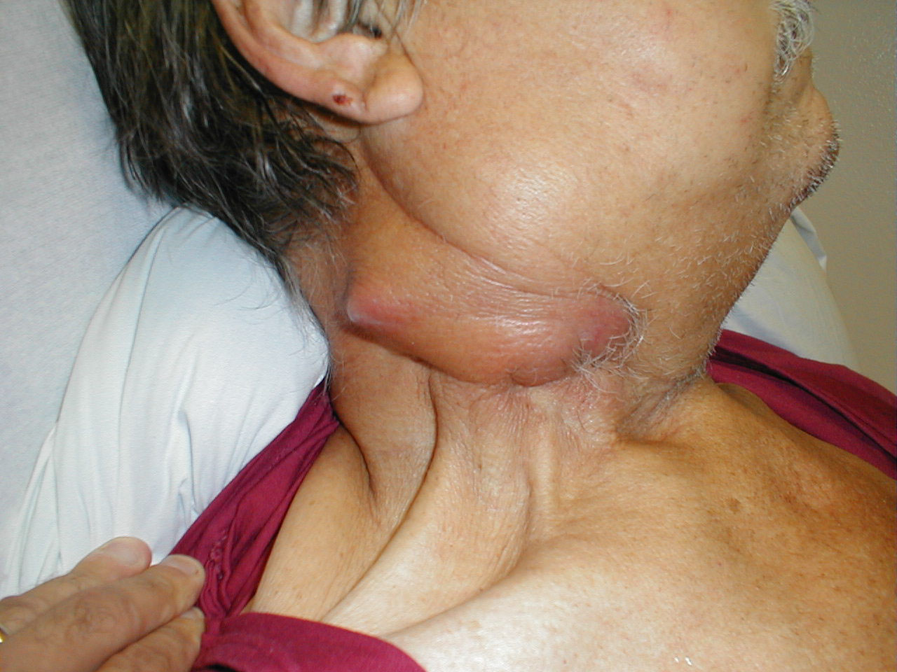
Cervical adenopathy: massive right side cervical adenopathy and facial asymmetry due to metastatic, intraoral squamous cell cancer. Images Courtesy of Charlie Goldberg, M.D., UCSD School of Medicine and VA Medical Center, San Diego, CA.
-
Cervical adenopathy: massive right side cervical adenopathy and facial asymmetry due to metastatic, intraoral squamous cell cancer. Images Courtesy of Charlie Goldberg, M.D., UCSD School of Medicine and VA Medical Center, San Diego, CA.
-
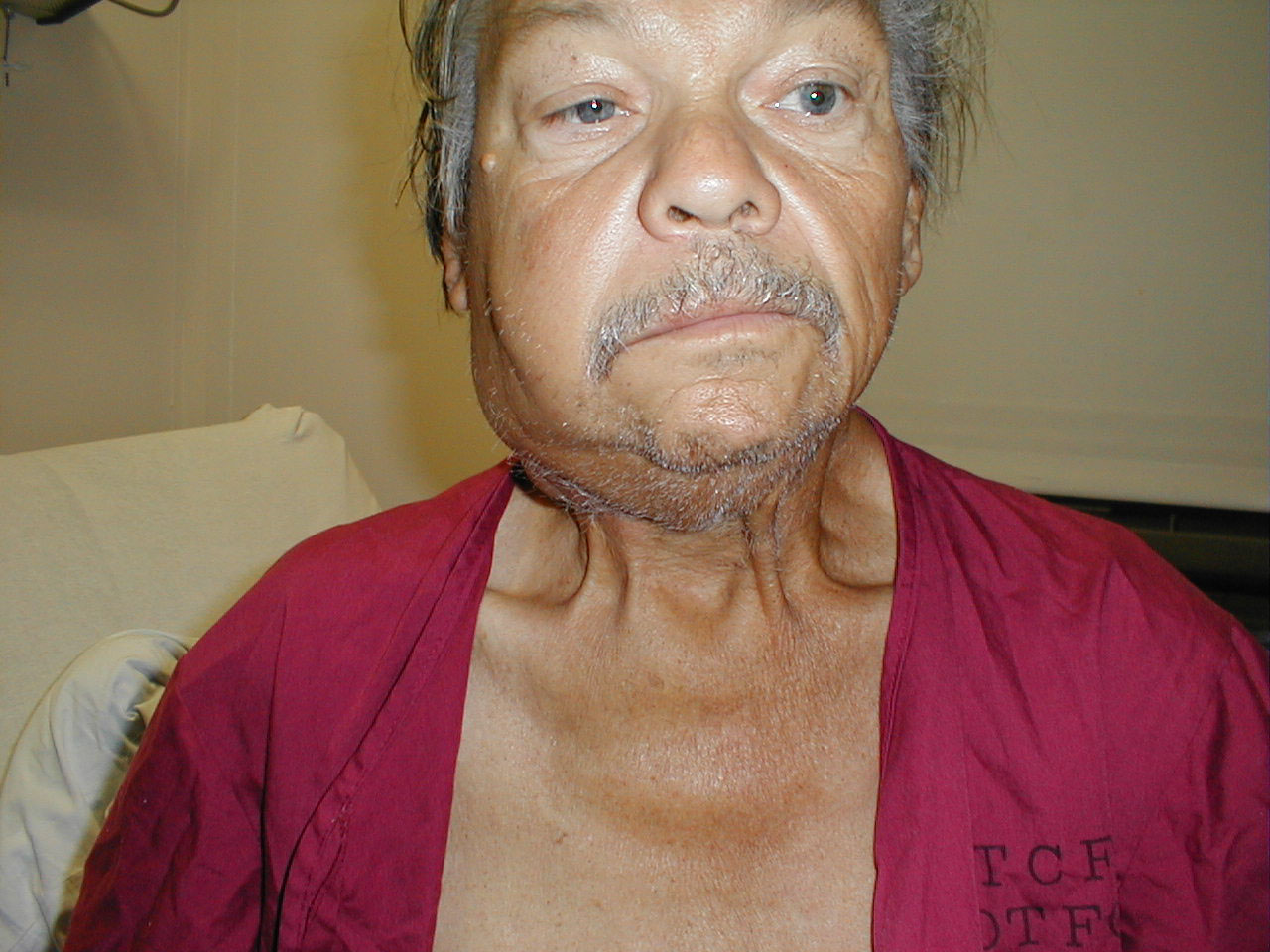
Cervical adenopathy: large right anterior cervical lymph node. Images Courtesy of Charlie Goldberg, M.D., UCSD School of Medicine and VA Medical Center, San Diego, CA.
-
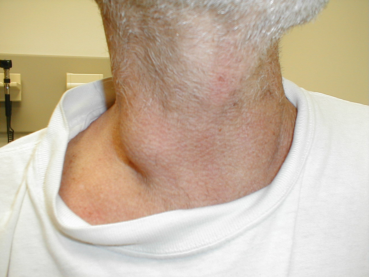
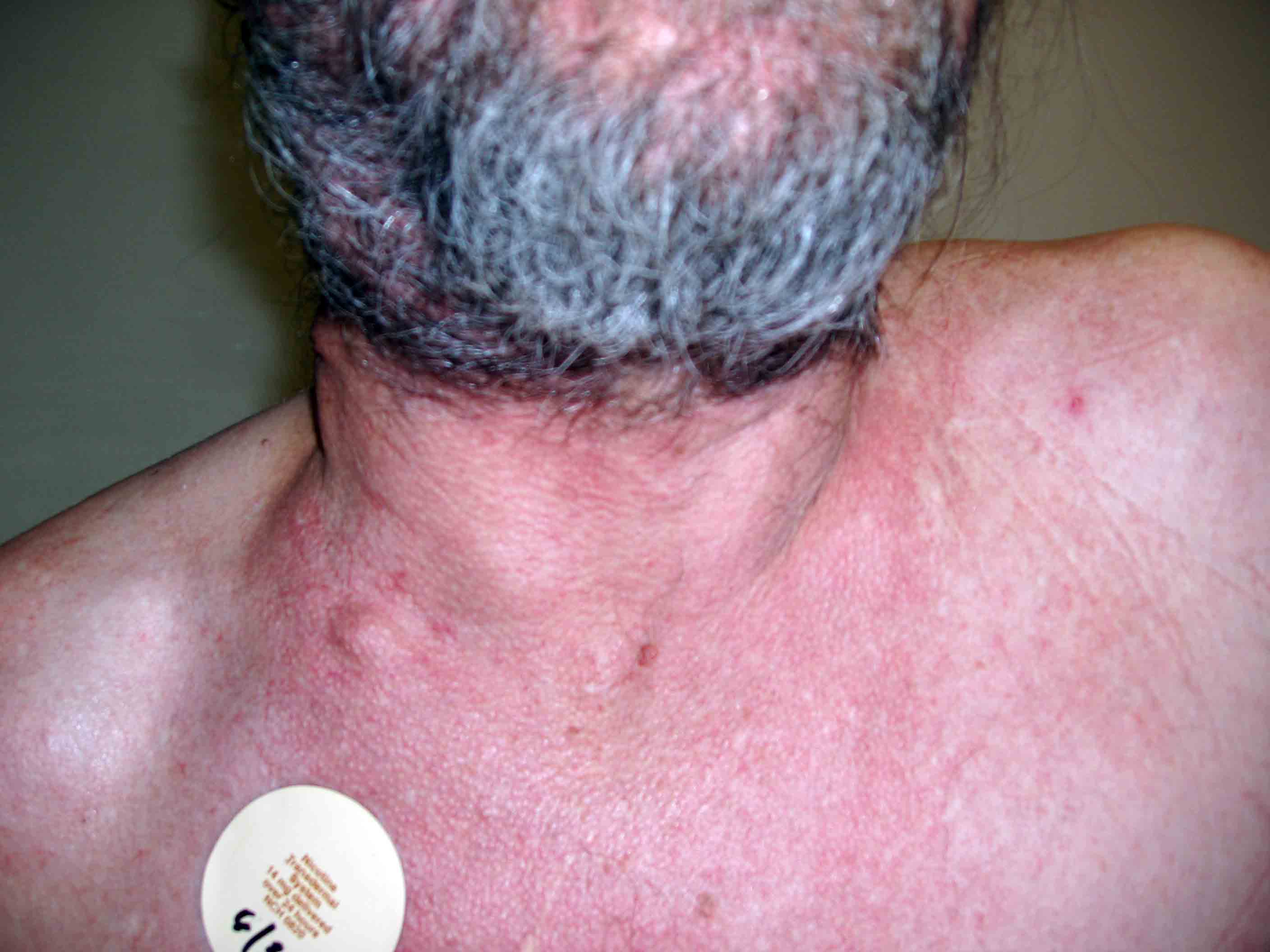
Cervical lymphadenopathy. Images Courtesy of Charlie Goldberg, M.D., UCSD School of Medicine and VA Medical Center, San Diego, CA.
-
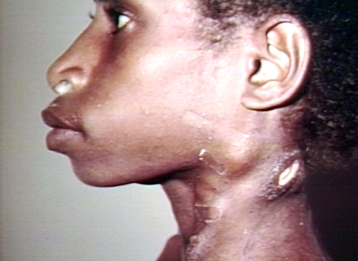
Cervical Adenopathy: multiple right sided cervical lymph nodes. Images Courtesy of Charlie Goldberg, M.D., UCSD School of Medicine and VA Medical Center, San Diego, CA.


Laboratory Findings
Complete Blood Count
- Elevated lactate dehydrogenase (LDH)
- Mild neutropenia
- Leukocytosis
- Elevated markers of inflammation and acute phase reactants (e.g. ESR, C-reactive protein, ferritin)
Imaging Findings
Ultrasonography
- Increased lymph node size.
CT
- Most nodes: 10 mm in short-axis
- Sub-mental and sub-mandibular: 15 mm
- Retropharyngeal: 8 mm
- Loss of fatty hilum
- Focal necrosis
- Cystic necrotic nodes
- Long-to-short axis ratio (>2cm - usually benign)
- The upper limit in size of a normal node varies with location.
Treatment
- There is no treatment for lymphadenopathy; the mainstay of therapy is treating the underlying condition.[2]
- For instance, infectious lymphadenopathy responds well to prompt treatment with antibiotics, and usually leads to a complete recovery. However, it may take months, for swelling to disappear. The amount of time to recovery depends on the cause.[3]
References
- ↑ King, D; Ramachandra, J; Yeomanson, D (2 January 2014). "Lymphadenopathy in children: refer or reassure?". Archives of Disease in Childhood: Education and Practice Edition. 99: 101–110. doi:10.1136/archdischild-2013-304443. PMID 24385291.
- ↑ 2.00 2.01 2.02 2.03 2.04 2.05 2.06 2.07 2.08 2.09 2.10 2.11 2.12 2.13 2.14 Mohseni S, Shojaiefard A, Khorgami Z, Alinejad S, Ghorbani A, Ghafouri A (2014). "Peripheral lymphadenopathy: approach and diagnostic tools". Iran J Med Sci. 39 (2 Suppl): 158–70. PMC 3993046. PMID 24753638.
- ↑ 3.00 3.01 3.02 3.03 3.04 3.05 3.06 3.07 3.08 3.09 3.10 3.11 3.12 3.13 3.14 3.15 Lymph node enlargment. Wikipedia. https://en.wikipedia.org/wiki/Lymph_node Accessed on May 9, 2016
- ↑ 4.0 4.1 Lymph node enlargment. Radiopedia. http://radiopaedia.org/articles/lymph-node-enlargement Accessed on May 9, 2016