Lymph node
|
WikiDoc Resources for Lymph node |
|
Articles |
|---|
|
Most recent articles on Lymph node |
|
Media |
|
Evidence Based Medicine |
|
Clinical Trials |
|
Ongoing Trials on Lymph node at Clinical Trials.gov Clinical Trials on Lymph node at Google
|
|
Guidelines / Policies / Govt |
|
US National Guidelines Clearinghouse on Lymph node
|
|
Books |
|
News |
|
Commentary |
|
Definitions |
|
Patient Resources / Community |
|
Patient resources on Lymph node Discussion groups on Lymph node Patient Handouts on Lymph node Directions to Hospitals Treating Lymph node Risk calculators and risk factors for Lymph node
|
|
Healthcare Provider Resources |
|
Causes & Risk Factors for Lymph node |
|
Continuing Medical Education (CME) |
|
International |
|
|
|
Business |
|
Experimental / Informatics |
Editor-In-Chief: C. Michael Gibson, M.S., M.D. [1]
Lymph nodes are components of the lymphatic system. They are sometimes informally called lymph glands but, as they do not secrete substances, such terminology is not entirely accurate. They are found mostly in the neck area.
Lymph nodes are filters or traps for foreign particles and contain white blood cells.
Function
Lymph nodes act as filters, with an internal honeycomb of reticular connective tissue filled with lymphocytes that collect and destroy bacteria and viruses. When the body is fighting an infection, lymphocytes multiply rapidly and produce a characteristic swelling of the lymph nodes.
Structure
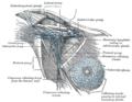
The lymph node is surrounded by a fibrous capsule, and inside the lymph node the fibrous capsule extends to form trabeculae. Thin reticular fibers form a supporting meshwork inside the node.
The concave side of the lymph node is called the hilum. The artery and vein attach at the hilum and allows blood to enter and leave the organ, respectively.
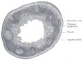
The parenchyma of the lymph node is divided into an outer cortex and an inner medulla.
Cortex
In the cortex, the subcapsular sinus drains to cortical sinusoids.
The outer cortex and inner cortex have very different properties:
| Location | Name/description | Predominant lymphocyte | Has nodules? |
| outer cortex | nodular cortex | B cells | yes |
| deep cortex | juxtamedullary cortex or paracortex | T cells | no |
The cortex is absent at the hilum.
It is made out of the fluid from the blood called plasma
Medulla
There are two named structures in the medulla:
- The medullary cords are cords of lymphatic tissue, and include plasma cells and T cells
- The medullary sinuses (or sinusoids) are vessel-like spaces separating the medullary cords. Lymph flows to the medullary sinuses from cortical sinuses, and into efferent lymphatic vessels. Medullary sinuses contain histiocytes (immobile macrophages) and reticular cells.
Shape and size
Human lymph nodes are bean-shaped and range in size from a few millimeters to about 1-2 cm in their normal state. They may become enlarged due to a tumor or infection. White blood cells are located within honeycomb structures of the lymph nodes. Lymph nodes are enlarged when the body is infected due to enhanced production of some cells and division of activated T and B cells. In some cases they may feel enlarged due to past infections; although one may be healthy, one may still feel them residually enlarged.
Lymphatic circulation
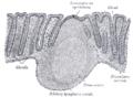
Lymph circulates to the lymph node via afferent lymphatic vessels and drains into the node just beneath the capsule in a space called the subcapsular sinus. The subcapsular sinus drains into trabecular sinuses and finally into medullary sinuses. The sinus space is criss-crossed by the pseudopods of macrophages which act to trap foreign particles and filter the lymph. The medullary sinuses converge at the hilum and lymph then leaves the lymph node via the efferent lymphatic vessel.
Lymphocytes, both B cells and T cells, constantly circulate through the lymph nodes. They enter the lymph node via the bloodstream and cross the wall of blood vessels by the process of diapedesis.
- The B cells migrate to the nodular cortex and medulla.
- The T cells migrate to the deep cortex.
When a lymphocyte recognizes an antigen, B cells become activated and migrate to germinal centers (by definition, a "secondary nodule" has a germinal center, while a "primary nodule" does not). When antibody-producing plasma cells are formed, they migrate to the medullary cords. Stimulation of the lymphocytes by antigens can accelerate the migration process to about 10 times normal, resulting in characteristic swelling of the lymph nodes.
The spleen and tonsils are large lymphoid organs that serve similar functions to lymph nodes, though the spleen filters blood cells rather than bacteria or viruses.
Distribution

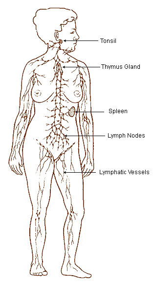
Humans have approximately 500-600 lymph nodes distributed throughout the body, with clusters found in the underarms, groin, neck, chest, and abdomen.
Lymph nodes of the human head and neck
- Cervical lymph nodes
- Anterior cervical: These nodes, both superficial and deep, lie above and beneath the sternocleidomastoid muscles. They drain the internal structures of the throat as well as part of the posterior pharynx, tonsils, and thyroid gland.
- Posterior cervical: These nodes extend in a line posterior to the sternocleidomastoids but in front of the trapezius, from the level of the Mastoid portion of the temporal bone to the clavicle. They are frequently enlarged during upper respiratory infections.
- Tonsillar: (sub mandibular) These nodes are located just below the angle of the mandible. They drain the tonsillar and posterior pharyngeal regions.
- Sub-mandibular: These nodes run along the underside of the jaw on either side. They drain the structures in the floor of the mouth.
- Sub-mental: These nodes are just below the chin. They drain the teeth and intra-oral cavity.
- Supraclavicular lymph nodes: These nodes are in the hollow above the clavicle, just lateral to where it joins the sternum. They drain a part of the thoracic cavity and abdomen. Virchow's node is a left supraclavicular lymph node which receives the lymph drainage from most of the body (especially the abdomen) via the thoracic duct and is thus an early site of metastasis for various malignancies.
Lymph nodes of the arm
These drain the whole of the arm, and are divided into two groups, superficial and deep. The superficial nodes are supplied by lymphatics which are present throughout the arm, but are particularly rich on the palm and flexor aspects of the digits.
- Superficial lymph glands of the arm:
- supratrochlear glands: Situated above the medial epicondyle of the humerus, medial to the basilic vein, they drain the C7 and C8 dermatomes.
- deltoideopectoral glands: Situated between the pectoralis major and deltoid muscles inferior to the clavicle.
- Deep lymph glands of the arm: These comprise the axillary glands, which are 20-30 individual glands and can be subdivided into:
- lateral glands
- anterior or pectoral glands
- posterior or subscapular glands
- central or intermediate glands
- medial or subclavicular glands
Lower limbs
Additional images
-
Lymphatic system
-
The human lymphatic system


-
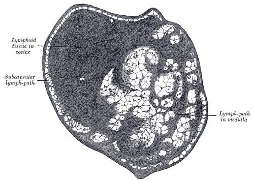
Section of small lymph node of rabbit. X 100.
-
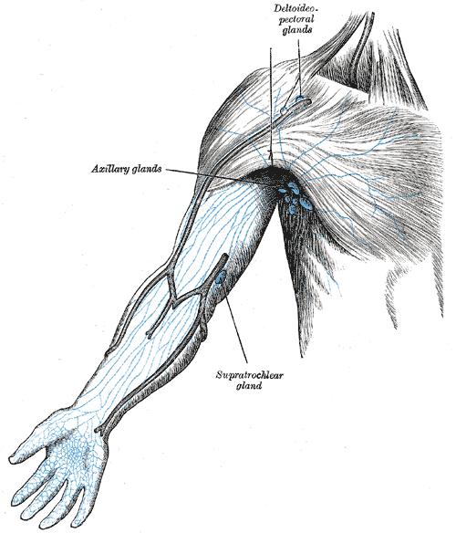
Lymphatics of the arm


-
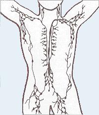
Lymphatics of the axillary region
-
Transverse section of human vermiform process. X 20.
-
Section of mucous membrane of human rectum. X 60.


Histopathological Findings of Lymph Node Diseases
Lymph node: Adenocarcinoma, metastatic to lymph node
{{#ev:youtube|AOXRSgsmAIc}}
Lymph node: Anthracosis
{{#ev:youtube|v4qufQkEIOY}}
Lymph node: Follicular hyperplasia
{{#ev:youtube|_ukg6FYkj8A}}
Lymph node: Metastatic undifferentiated large cell carcinoma
{{#ev:youtube|JgzZ390-UWI}}
Lymph node: Lymphocyte depleted lymph node
{{#ev:youtube|OzHJkkFnlvU}}
Lymph node: Mycobacterium avium-intracellulare complex (MAC)
{{#ev:youtube|MWHD80xx3SM}}
Lymph node: Tuberculosis
{{#ev:youtube|pa8ESYPZ6sk}}
Lymphogranuloma
{{#ev:youtube|F5sD6FgjLmA}}
Lymph node: Metastatic breast carcinoma
{{#ev:youtube|8-8V2ZcTiXQ}}
Lymph node: Acute Lymphadenitis
{{#ev:youtube|it0lwR3Fls0}}
Lymph node: Brucellosis
{{#ev:youtube|BGAb9IGjotA}}
Lymph node: Toxoplasmosis
{{#ev:youtube|fyU-LEyV0aQ}}
Lymph node: Tularemia
{{#ev:youtube|E9sT4X36etA}}
See also
External links
- Immunology Lecture 4, Biology 328 at Western Kentucky University
- Lymph Nodes
- Histology image: 07101loa – Histology Learning System at Boston University
da:Lymfeknude de:Lymphknoten eo:Limfaj ganglioj fa:گره لنفی ia:Glandula lymphatic it:Linfonodo lt:Limfmazgis nl:Lymfeklier fi:Imusolmuke sv:Lymfknuta uk:Лімфатичні вузли