Intracerebral hemorrhage CT: Difference between revisions
(Created page with "__NOTOC__ {{Intracerebral hemorrhage}} {{CMG}}; {{AE}} {{SaraM}} ==Overview== ==Ct scan== ==References== {{reflist|2}} Category:Neurology Category:Cardiology C...") |
(→CT) |
||
| (3 intermediate revisions by the same user not shown) | |||
| Line 5: | Line 5: | ||
==Overview== | ==Overview== | ||
CT is very sensitive for identifying acute hemorrhage and is considered the '''gold standard'''.<ref name=Fiebach>Fiebach JB, Schellinger PD, Gass A, Kucinski T, Siebler M, Villringer A, Olkers P, Hirsch JG, Heiland S, Wilde P, Jansen O, Röther J, Hacke W, Sartor K; Kompetenznetzwerk Schlaganfall B5. Stroke magnetic resonance imaging is accurate in hyperacute intracerebral hemorrhage: a multicenter study on the validity of stroke imaging. Stroke. 2004;35:502– 506. doi: 10.1161/01.STR.0000114203.75678.8</ref><ref name=Chalela>Chalela JA, Kidwell CS, Nentwich LM, Luby M, Butman JA, Demchuk AM, Hill MD, Patronas N, Latour L, Warach S. Magnetic resonance imaging and computed tomography in emergency assessment of patients with suspected acute stroke: a prospective comparison. Lancet. 2007;369:293–298. doi: 10.1016/S0140-6736(07)60151-2.</ref> | |||
==CT== | |||
Noncontrast cranial CT is very sensitive for identifying acute hemorrhage and is considered the '''gold standard'''.<ref name=Fiebach>Fiebach JB, Schellinger PD, Gass A, Kucinski T, Siebler M, Villringer A, Olkers P, Hirsch JG, Heiland S, Wilde P, Jansen O, Röther J, Hacke W, Sartor K; Kompetenznetzwerk Schlaganfall B5. Stroke magnetic resonance imaging is accurate in hyperacute intracerebral hemorrhage: a multicenter study on the validity of stroke imaging. Stroke. 2004;35:502– 506. doi: 10.1161/01.STR.0000114203.75678.8</ref><ref name=Chalela>Chalela JA, Kidwell CS, Nentwich LM, Luby M, Butman JA, Demchuk AM, Hill MD, Patronas N, Latour L, Warach S. Magnetic resonance imaging and computed tomography in emergency assessment of patients with suspected acute stroke: a prospective comparison. Lancet. 2007;369:293–298. doi: 10.1016/S0140-6736(07)60151-2.</ref><ref name="pmid18275927">{{cite journal| author=Kidwell CS, Wintermark M| title=Imaging of intracranial haemorrhage. | journal=Lancet Neurol | year= 2008 | volume= 7 | issue= 3 | pages= 256-67 | pmid=18275927 | doi=10.1016/S1474-4422(08)70041-3 | pmc= | url=https://www.ncbi.nlm.nih.gov/entrez/eutils/elink.fcgi?dbfrom=pubmed&tool=sumsearch.org/cite&retmode=ref&cmd=prlinks&id=18275927 }} </ref> | |||
== | The following information can be defined by CT scan: | ||
*The size and location of the [[hematoma]] | |||
*Ventricular extension of the [[hematoma]] | |||
*Presence of [[edema]] | |||
*Brain shifts and [[herniation]] | |||
{| style="border: 0px; font-size: 90%; margin: 3px;" align=center | |||
|+ | |||
! style="background: #4479BA; width: 220px;" | {{fontcolor|#FFF|Type of hemorrhage}} | |||
! style="background: #4479BA; width: 550px;" | {{fontcolor|#FFF|Radiologic criteria}} | |||
|- | |||
| style="padding: 5px 5px; background: #F5F5F5;" |'''Hyperacute | |||
| style="padding: 5px 5px; background: #F5F5F5;" | | |||
*Hyperdense | |||
|- | |||
| style="padding: 5px 5px; background: #F5F5F5;" |'''After one week | |||
| style="padding: 5px 5px; background: #F5F5F5;" | | |||
*Isodense | |||
*Ring enhancemen | |||
|- | |||
| style="padding: 5px 5px; background: #F5F5F5;" | '''Chronic | |||
| style="padding: 5px 5px; background: #F5F5F5;" | | |||
*Hypodense | |||
|- | |||
| style="padding: 5px 5px; background: #F5F5F5;" |'''Hemorrhagic transformation of a cerebral infarct | |||
| style="padding: 5px 5px; background: #F5F5F5;" | | |||
*Patchy appearance of the hyperdensity within a larger area of low attenuation | |||
*Wedge-shaped abnormality that extends to the cortex | |||
|} | |||
==Images== | |||
The following are images associated with intracerebral haemorrhage.<ref name=HS-radiopaedia> Intracerebral Hemotrrhage https://radiopaedia.org/cases/intracerebral-haemorrhage-2 Accessed on November 9, 2016</ref> | |||
<div align="center"> | |||
<gallery heights="175" widths="175"> | |||
Image:Intracerebral haemorrhage.jpg|Large hemorrhagic focus in the left cerebral hemisphere that extends to the infratentorial region with a significant mass effect | |||
Image:Hypertensive intracerebral haemorrhage.jpg|Large hemorrhagic focus in the left cerebral hemisphere that extends to the infratentorial region with a significant mass effect | |||
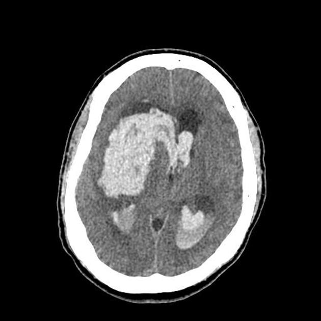
Image:Coagulopathy related intracerebral haemorrhage.jpg|Multiple descrete bilateral supratentorial intraparenchymal cerebral haemorrhage | |||
Image:Intracerebral parenchymal haemorrhage.jpeg|Very large intracerebral haemorrhage on the left extends to involve the ventricles. It exerts marked mass effect | |||
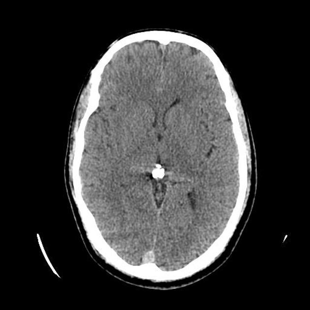
Image:Haemorrhagic stroke- basal ganglia.jpg|Hemorrhagic stroke in the left basal ganglia | |||
</gallery> | |||
</div> | |||
==References== | ==References== | ||
Latest revision as of 03:04, 30 November 2016
|
Intracerebral hemorrhage Microchapters |
|
Diagnosis |
|---|
|
Treatment |
|
AHA/ASA Guidelines for the Management of Spontaneous Intracerebral Hemorrhage (2015) |
|
AHA/ASA Guideline Recommendation for the Primary Prevention of Stroke (2014) |
|
Case Studies |
|
Intracerebral hemorrhage CT On the Web |
|
American Roentgen Ray Society Images of Intracerebral hemorrhage CT |
|
Risk calculators and risk factors for Intracerebral hemorrhage CT |
Editor-In-Chief: C. Michael Gibson, M.S., M.D. [1]; Associate Editor(s)-in-Chief: Sara Mehrsefat, M.D. [2]
Overview
CT is very sensitive for identifying acute hemorrhage and is considered the gold standard.[1][2]
CT
Noncontrast cranial CT is very sensitive for identifying acute hemorrhage and is considered the gold standard.[1][2][3]
The following information can be defined by CT scan:
- The size and location of the hematoma
- Ventricular extension of the hematoma
- Presence of edema
- Brain shifts and herniation
| Type of hemorrhage | Radiologic criteria |
|---|---|
| Hyperacute |
|
| After one week |
|
| Chronic |
|
| Hemorrhagic transformation of a cerebral infarct |
|
Images
The following are images associated with intracerebral haemorrhage.[4]
-
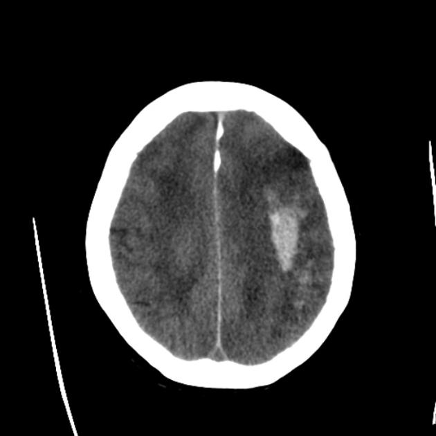
Large hemorrhagic focus in the left cerebral hemisphere that extends to the infratentorial region with a significant mass effect
-
Large hemorrhagic focus in the left cerebral hemisphere that extends to the infratentorial region with a significant mass effect
-
Multiple descrete bilateral supratentorial intraparenchymal cerebral haemorrhage
-
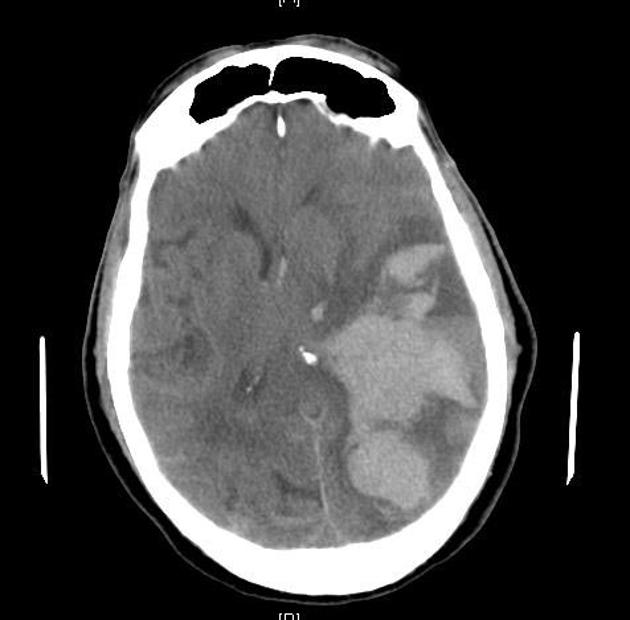
Very large intracerebral haemorrhage on the left extends to involve the ventricles. It exerts marked mass effect
-
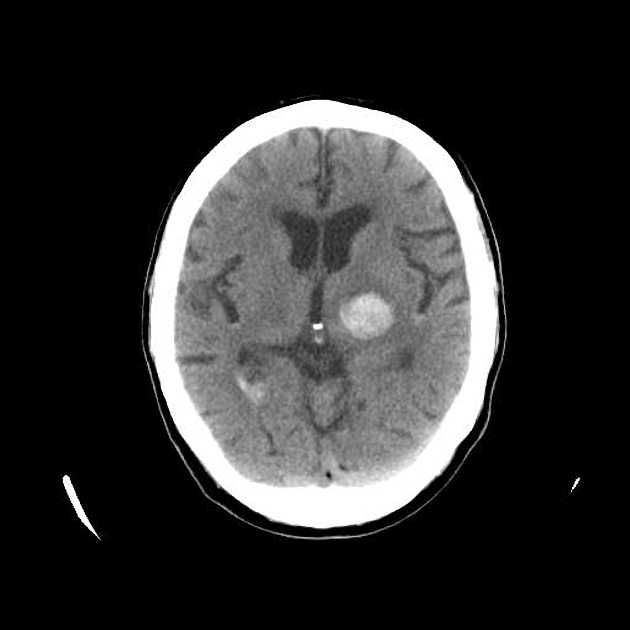
Hemorrhagic stroke in the left basal ganglia


References
- ↑ 1.0 1.1 Fiebach JB, Schellinger PD, Gass A, Kucinski T, Siebler M, Villringer A, Olkers P, Hirsch JG, Heiland S, Wilde P, Jansen O, Röther J, Hacke W, Sartor K; Kompetenznetzwerk Schlaganfall B5. Stroke magnetic resonance imaging is accurate in hyperacute intracerebral hemorrhage: a multicenter study on the validity of stroke imaging. Stroke. 2004;35:502– 506. doi: 10.1161/01.STR.0000114203.75678.8
- ↑ 2.0 2.1 Chalela JA, Kidwell CS, Nentwich LM, Luby M, Butman JA, Demchuk AM, Hill MD, Patronas N, Latour L, Warach S. Magnetic resonance imaging and computed tomography in emergency assessment of patients with suspected acute stroke: a prospective comparison. Lancet. 2007;369:293–298. doi: 10.1016/S0140-6736(07)60151-2.
- ↑ Kidwell CS, Wintermark M (2008). "Imaging of intracranial haemorrhage". Lancet Neurol. 7 (3): 256–67. doi:10.1016/S1474-4422(08)70041-3. PMID 18275927.
- ↑ Intracerebral Hemotrrhage https://radiopaedia.org/cases/intracerebral-haemorrhage-2 Accessed on November 9, 2016