Hymenolepis infection
Editor-In-Chief: C. Michael Gibson, M.S., M.D. [1]
Related Key Words and Synonyms: Dwarf tapeworm, Hymenolepiasis.
Pathophysiology & Etiology
Etiologic agent:
Hymenolepiasis is caused by two cestodes (tapeworm) species, Hymenolepis nana (the dwarf tapeworm, adults measuring 15 to 40 mm in length) and Hymenolepis dimnuta (rat tapeworm, adults measuring 20 to 60 cm in length). Hymenolepis diminuta is a cestode of rodents infrequently seen in humans and frequently found in rodents.
Life cycle:
- Hymenolepis nana
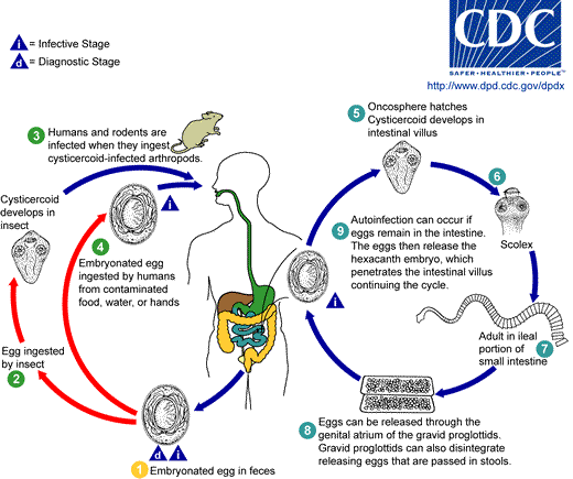
Eggs of Hymenolepis nana are immediately infective when passed with the stool and cannot survive more than 10 days in the external environment 1. When eggs are ingested by an arthropod intermediate host 2 (various species of beetles and fleas may serve as intermediate hosts), they develop into cysticercoids, which can infect humans or rodents upon ingestion 3 and develop into adults in the small intestine. A morphologically identical variant, H. nana var. fraterna, infects rodents and uses arthropods as intermediate hosts. When eggs are ingested 4 (in contaminated food or water or from hands contaminated with feces), the oncospheres contained in the eggs are released. The oncospheres (hexacanth larvae) penetrate the intestinal villus and develop into cysticercoid larvae 5. Upon rupture of the villus, the cysticercoids return to the intestinal lumen, evaginate their scoleces 6, attach to the intestinal mucosa and develop into adults that reside in the ileal portion of the small intestine producing gravid proglottids 7. Eggs are passed in the stool when released from proglottids through its genital atrium or when proglottids disintegrate in the small intestine 8. An alternate mode of infection consists of internal autoinfection, where the eggs release their hexacanth embryo, which penetrates the villus continuing the infective cycle without passage through the external environment 9. The life span of adult worms is 4 to 6 weeks, but internal autoinfection allows the infection to persist for years.
- Hymenolepis diminuta
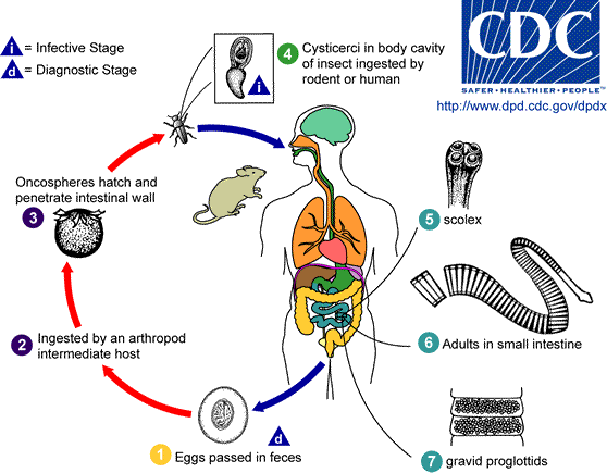
Eggs of Hymenolepis diminuta are passed out in the feces of the infected definitive host (rodents, man) 1. The mature eggs are ingested by an intermediate host (various arthropod adults or larvae) 2, and oncospheres are released from the eggs and penetrate the intestinal wall of the host 3, which develop into cysticercoid larvae 4. Species from the genus Tribolium are common intermediate hosts for H. diminuta. The cysticercoid larvae persist through the arthropod's morphogenesis to adulthood. H. diminuta infection is acquired by the mammalian host after ingestion of an intermediate host carrying the cysticercoid larvae . Humans can be accidentally infected through the ingestion of insects in precooked cereals, or other food items, and directly from the environment (e.g., oral exploration of the environment by children). After ingestion, the tissue of the infected arthropod is digested releasing the cysticercoid larvae in the stomach and small intestine. Eversion of the scoleces 5 occurs shortly after the cysticercoid larvae are released. Using the four suckers on the scolex, the parasite attaches to the small intestine wall. Maturation of the parasites occurs within 20 days and the adult worms can reach an average of 30 cm in length 6. Eggs are released in the small intestine from gravid proglottids 7 that disintegrate after breaking off from the adult worms. The eggs are expelled to the environment in the mammalian host's feces 1.
Diagnosis
The diagnosis depends on the demonstration of eggs in stool specimens. Concentration techniques and repeated examinations will increase the likelihood of detecting light infections.
Differential Diagnosis
History and Symptoms
Most people who are infected do not have any symptoms. Those who have symptoms may experience nausea, weakness, loss of appetite, diarrhea, and abdominal pain. Young children, especially those with a heavy infection, may develop a headache, itchy bottom, or have difficulty sleeping. Sometimes infection is misdiagnosed as a pinworm infection.
Contrary to popular belief, a tapeworm infection does not generally cause weight loss. You cannot feel the tapeworm inside your body.
Laboratory Findings
Microscopy:
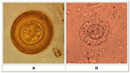
A: Egg of Hymenolepis diminuta. These eggs are round or slightly oval, size 70 to 86 µm by 60 to 80 µm, with a striated outer membrane and a thin inner membrane. The space between the membranes is smooth or faintly granular. The oncosphere has six hooks (of which at least four are visible at this level of focus). Image contributed by Georgia Department of Public Health.
B: Egg of Hymenolepis nana. These eggs are oval and smaller than those of H. diminuta, their size being 30 to 55 µm. On the inner membrane are two poles, from which 4 to 8 polar filaments spread out between the two membranes. The oncosphere has six hooks (seen as dark lines at 8 o'clock). Image contributed by Georgia Department of Public Health.
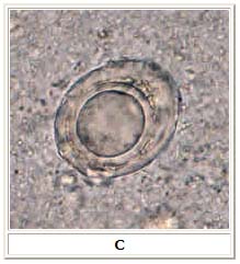
C: Artifact resembling a H. nana egg. However, no hooks and no polar filaments are visible in the artifact.
Macroscopic:
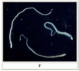
F: Three adult Hymenolepis nana tapeworms. Each tapeworm (length: 15 to 40 mm) has a small, rounded scolex at the anterior end, and proglottids can be distinguished at the posterior, wider end. Image contributed by the Georgia Division of Public Health.
Risk Stratification and Prognosis
- Infection with the dwarf tapeworm is generally not serious. However, prolonged infection can lead to more severe symptoms; therefore, medical attention is needed to eliminate the tapeworm.
- Eggs are infectious (meaning they can re-infect you or infect others) immediately after being shed in feces.
Treatment
Acute Pharmacotherapies
A prescription drug called praziquantel is given. The medication causes the tapeworm to dissolve within the intestines. Praziquantel is generally well tolerated. Sometimes more than one treatment is necessary.
Primary Prevention
- Wash hands with soap and water after using the toilet, and before handling food.
- If you work in a childcare center where you change diapers, be sure to wash your hands thoroughly with plenty of soap and warm water after every diaper change, even if you wear gloves.
- When traveling in countries where food is likely to be contaminated, wash, peel or cook all raw vegetables and fruits with safe water before eating.
References
- http://www.cdc.gov/ncidod/dpd/parasites/hymenolepis/default.htm
- http://www.dpd.cdc.gov/dpdx/HTML/Hymenolepiasis.htm
Acknowledgements
The content on this page was first contributed by: C. Michael Gibson, M.S., M.D.