Hydronephrosis pathophysiology: Difference between revisions
| Line 32: | Line 32: | ||
* Urinary tract infection | * Urinary tract infection | ||
*Congenital malformation | *Congenital malformation | ||
*Tumor | *Tumor<ref name="pmid29875948">{{cite journal |vauthors=Rachidi SA, Zeriouel A |title=[Renal tumor or pseudotumoral xanthogranulomatous pyelonephritis] |language=French |journal=Pan Afr Med J |volume=29 |issue= |pages=67 |date=2018 |pmid=29875948 |pmc=5987130 |doi=10.11604/pamj.2018.29.67.14611 |url=}}</ref> | ||
*Pregnancy | *Pregnancy | ||
* Scarring of tissues( From injury or previous surgery) | * Scarring of tissues( From injury or previous surgery) | ||
Revision as of 19:06, 23 July 2018
| https://https://www.youtube.com/watch?v=mi7XtyHwVHk%7C350}} |
|
Hydronephrosis Microchapters |
|
Diagnosis |
|---|
|
Treatment |
|
Case Studies |
|
Hydronephrosis pathophysiology On the Web |
|
American Roentgen Ray Society Images of Hydronephrosis pathophysiology |
|
Risk calculators and risk factors for Hydronephrosis pathophysiology |
Editor-In-Chief: C. Michael Gibson, M.S., M.D. [1]; Associate Editor(s)-in-Chief: Vindhya BellamKonda, M.B.B.S [2]
Overview
Hydronephrosis can result from anatomic or functional processes interrupting the flow of urine. This interruption can occur anywhere along the urinary tract from the kidneys to the urethral meatus. The rise in ureteral pressure leads to marked changes in glomerular filtration, tubular function, and renal blood flow. The glomerular filtration rate (GFR) declines significantly within hours following acute obstruction. This significant decline of GFR can persist for weeks after relief of obstruction. In addition, renal tubular ability to transport sodium, potassium, and protons and concentrate and to dilute the urine is severely impaired.
Pathophysiology
- Hydronephrosis can result from anatomic or functional processes interrupting the flow of urine.
- This interruption can occur anywhere along the urinary tract from the kidneys to the urethral meatus. The rise in ureteral pressure leads to marked changes in glomerular filtration, tubular function, and renal blood flow.
- The glomerular filtration rate (GFR) declines significantly within hours following acute obstruction. This significant decline of GFR can persist for weeks after relief of obstruction. In addition, renal tubular ability to transport sodium, potassium, and protons and concentrate and to dilute the urine is severely impaired.
- The obstruction may be either partial or complete and can occur anywhere from the urethral meatus to the calyces of the renal pelvis.
- The obstruction may arise from either inside or outside the urinary tract or may come from the wall of the urinary tract itself.
- Intrinsic obstructions (those that occur within the tract) include blood clots, stones, sloughed papilla along with tumours of the kidney, ureter and bladder. Extrinsic obstructions (those that are caused by factors outside of the urinary tract) include pelvic or abdominal tumours or masses, retroperitoneal fibrosis or neurological deficits.
- Strictures of the ureters (congenital or acquired), neuromuscular dysfunctions or schistosomiasis are other causes which originate from the wall of the urinary tract.[1][2]
Genetics
- Hydronephrosis is transmitted in autosomal dominant pattern.[3]
- Evidence for genetic heterogeneity in hereditary hydronephrosis caused by pelvic-ureteric junction obstruction with one locus assigned to chromosome 6p has been found.[4][5]
Associated Conditions
- Nephrolithiasis[6]
- Urinary tract infection
- Congenital malformation
- Tumor[7]
- Pregnancy
- Scarring of tissues( From injury or previous surgery)
Gross Pathology
- On gross pathology, the following features may be see in hydronephrosis.
- Renal papillary compression
Microscopic Pathology
- On microscopic histopathological analysis, [feature1], [feature2], and [feature3] are characteristic findings of [disease name].
-
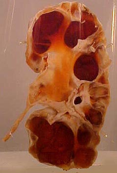
Specimen of a kidney that has undergone extensive dilation due to hydronephrosis. Note the extensive atrophy and thinning of the renal cortex.

References
- ↑ Krajewski W, Wojciechowska J, Dembowski J, Zdrojowy R, Szydełko T (August 2017). "Hydronephrosis in the course of ureteropelvic junction obstruction: An underestimated problem? Current opinions on the pathogenesis, diagnosis and treatment". Adv Clin Exp Med. 26 (5): 857–864. PMID 29068584.
- ↑ Vercellini P, Buggio L, Somigliana E (December 2017). "Role of medical therapy in the management of deep rectovaginal endometriosis". Fertil. Steril. 108 (6): 913–930. doi:10.1016/j.fertnstert.2017.08.038. PMID 29202965.
- ↑ Izquierdo L, Porteous M, Paramo PG, Connor JM (July 1992). "Evidence for genetic heterogeneity in hereditary hydronephrosis caused by pelvi-ureteric junction obstruction, with one locus assigned to chromosome 6p". Hum. Genet. 89 (5): 557–60. PMID 1634233.
- ↑ Fryns JP, Kleczkowska A, Moerman P, Vandenberghe K (June 1993). "Hereditary hydronephrosis and the short arm of chromosome 6". Hum. Genet. 91 (5): 514–5. PMID 8357406.
- ↑ Neas KR, Chia N, Clarke M, Peters G, Adès LC (July 2003). "A case of partial trisomy 4p syndrome presenting as severe hydronephrosis in utero". Clin. Dysmorphol. 12 (3): 179–81. doi:10.1097/01.mcd.0000072162.33788.8d. PMID 14564156.
- ↑ Wojciechowska M, Bieniaś B, Sobieszczańska-Droździel A, Wieczorkiewicz-Płaza A, Beń-Skowronek I, Sikora P (April 2018). "[Recurrent urolithiasis as a symptom of primary hyperparathyroidism in a 16-year-old boy]". Pol. Merkur. Lekarski (in Polish). 44 (262): 208–210. PMID 29775451.
- ↑ Rachidi SA, Zeriouel A (2018). "[Renal tumor or pseudotumoral xanthogranulomatous pyelonephritis]". Pan Afr Med J (in French). 29: 67. doi:10.11604/pamj.2018.29.67.14611. PMC 5987130. PMID 29875948.