Hydrocephalus MRI
|
Hydrocephalus Microchapters |
|
Diagnosis |
|---|
|
Treatment |
|
Case Studies |
|
Hydrocephalus MRI On the Web |
|
American Roentgen Ray Society Images of Hydrocephalus MRI |
Editor-In-Chief: C. Michael Gibson, M.S., M.D. [1];
Associate Editor-In-Chief: Cafer Zorkun M.D., PhD.,Kalsang Dolma, M.B.B.S.[2]
MRI
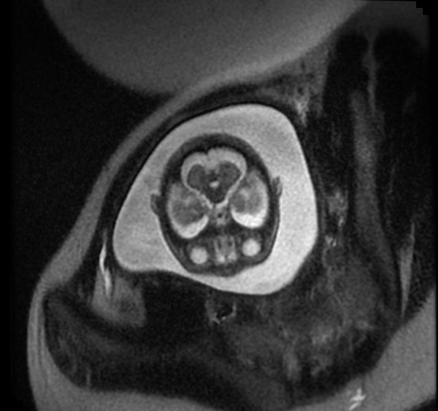
MRI gives the highest resolution of brain. It gives better picture of posterior fossa. It can evaluate chiari malformation and aqueductal stenosis
Aqueductal Stenosis
-
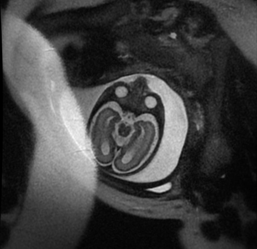
Fetal MRI: Aqueductal stenosis
-
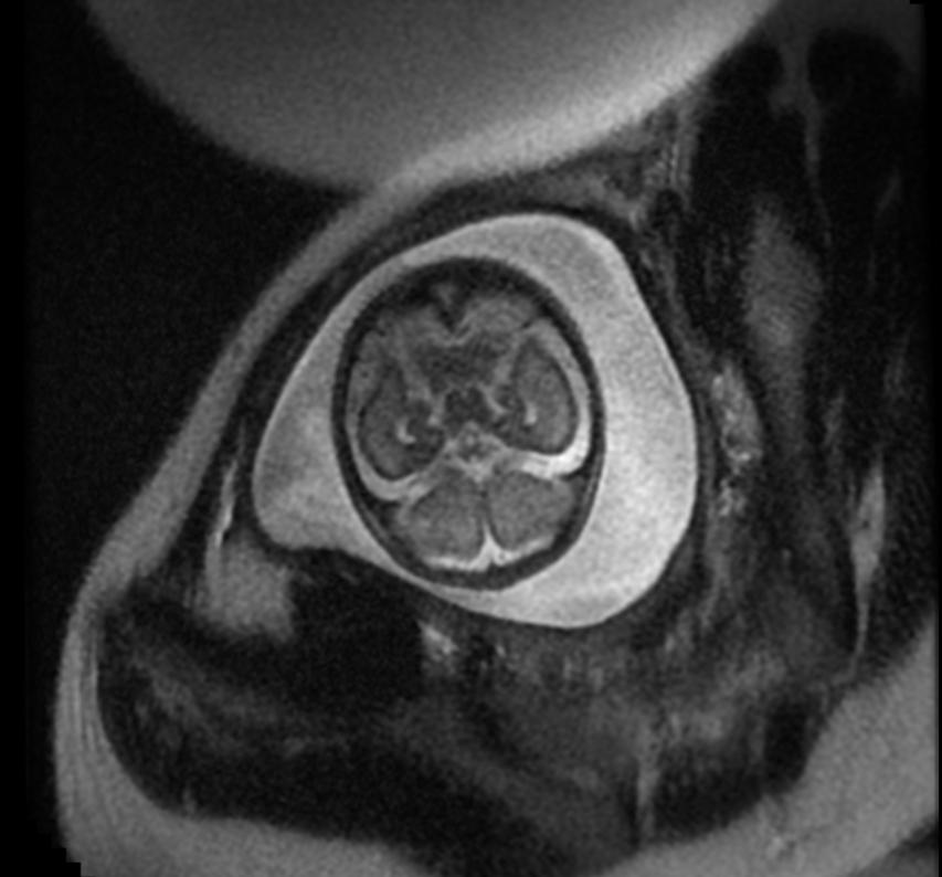
Fetal MRI: Aqueductal stenosis
-
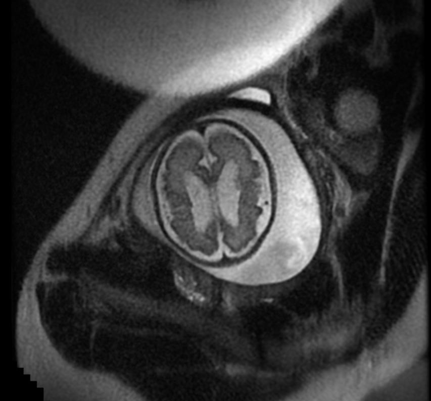
Fetal MRI: Aqueductal stenosis
-
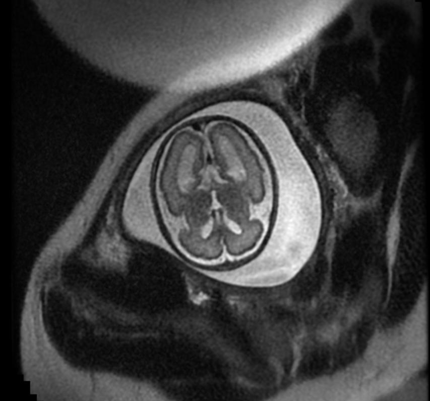
Fetal MRI: Aqueductal stenosis
-
Fetal MRI: Aqueductal stenosis


