Hydrocephalus MRI
|
Hydrocephalus Microchapters |
|
Diagnosis |
|---|
|
Treatment |
|
Case Studies |
|
Hydrocephalus MRI On the Web |
|
American Roentgen Ray Society Images of Hydrocephalus MRI |
Editor-In-Chief: C. Michael Gibson, M.S., M.D. [1]; Associate Editor-In-Chief: Cafer Zorkun M.D., PhD.,Kalsang Dolma, M.B.B.S.[2]
MRI
MRI gives the highest resolution images of the brain. It provides better pictures of the posterior fossa. It can be used to evaluate chiari malformation and aqueductal stenosis.
Marked Hyrocephalus


Aqueductal Stenosis
-
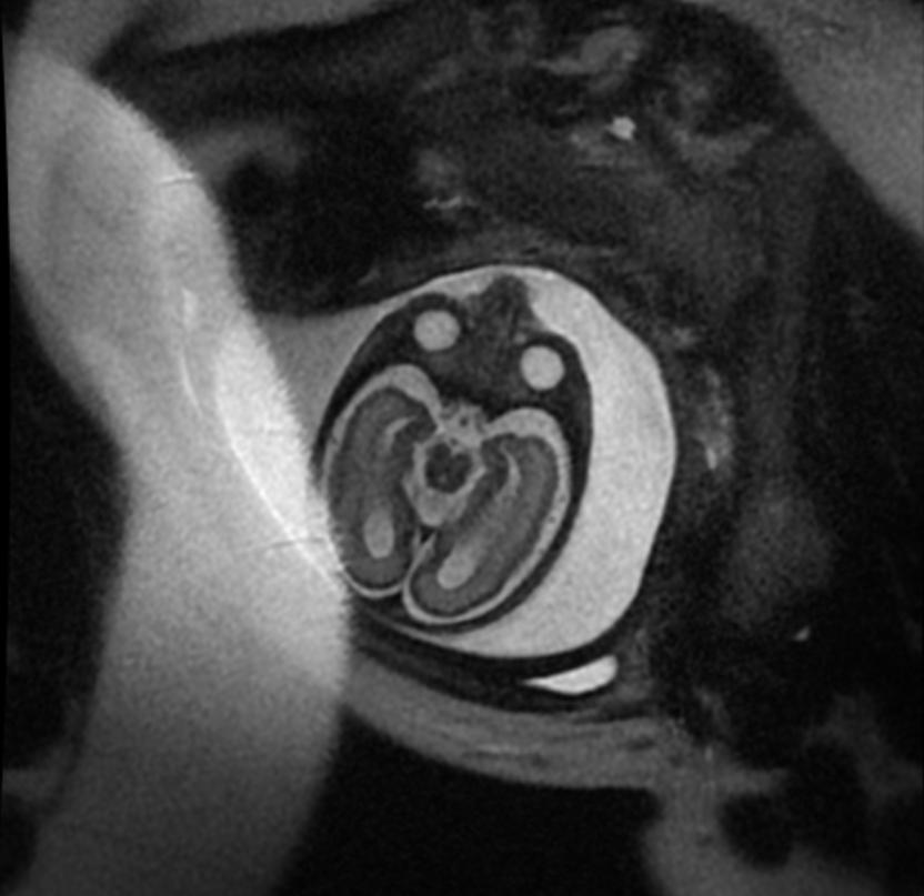
Fetal MRI: Aqueductal stenosis
-
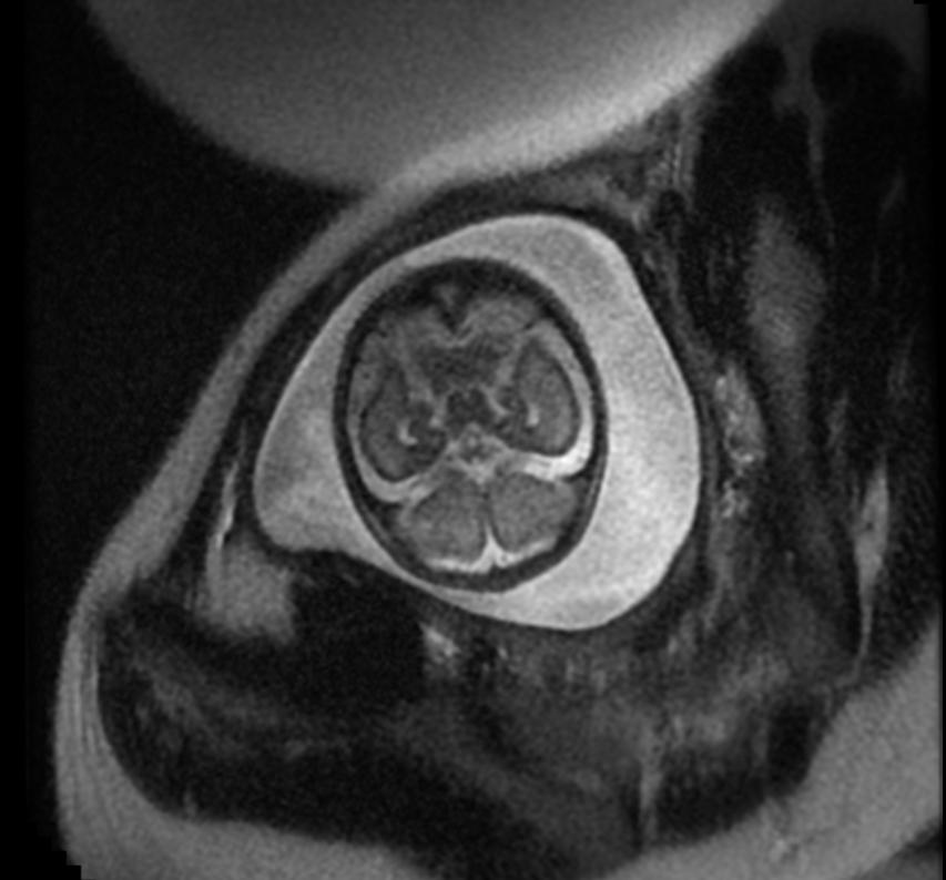
Fetal MRI: Aqueductal stenosis
-
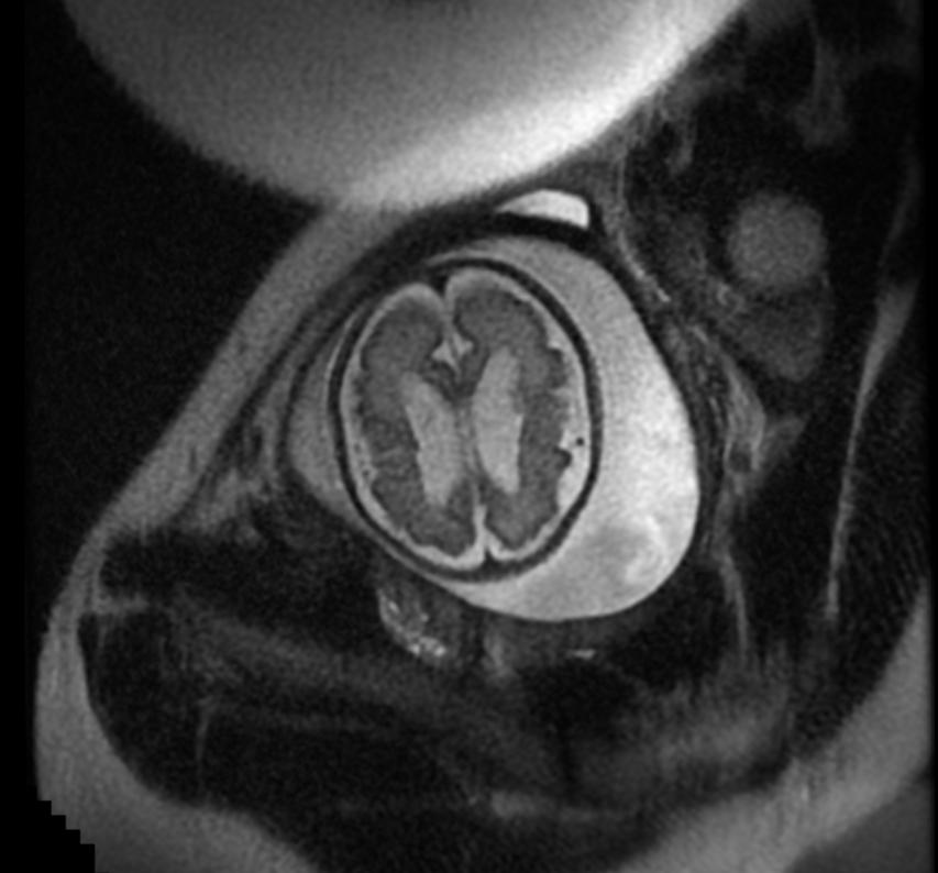
Fetal MRI: Aqueductal stenosis
-
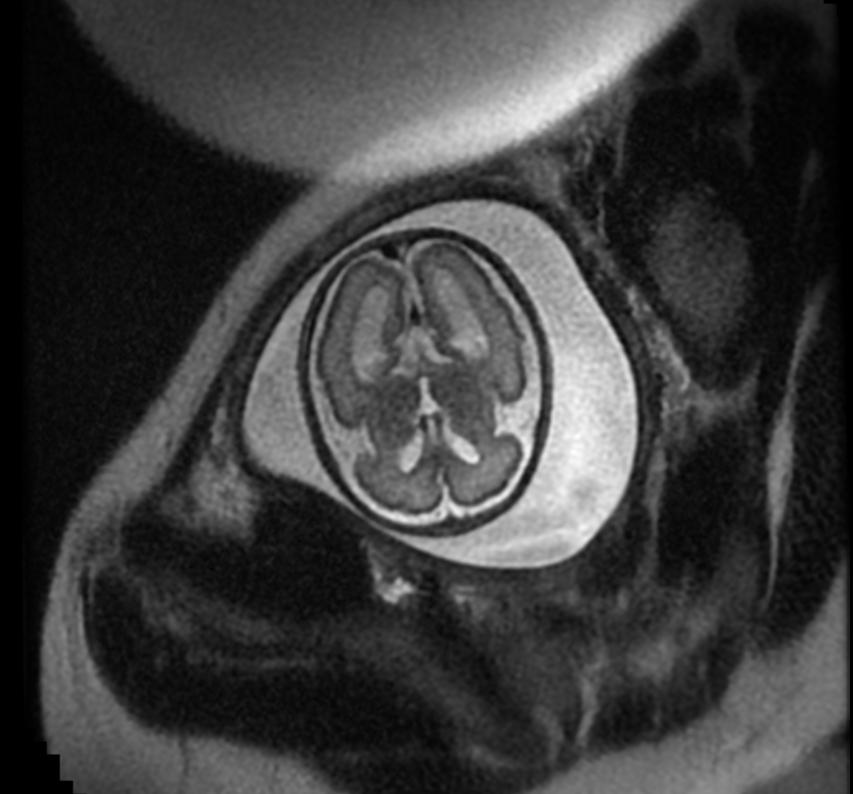
Fetal MRI: Aqueductal stenosis
-
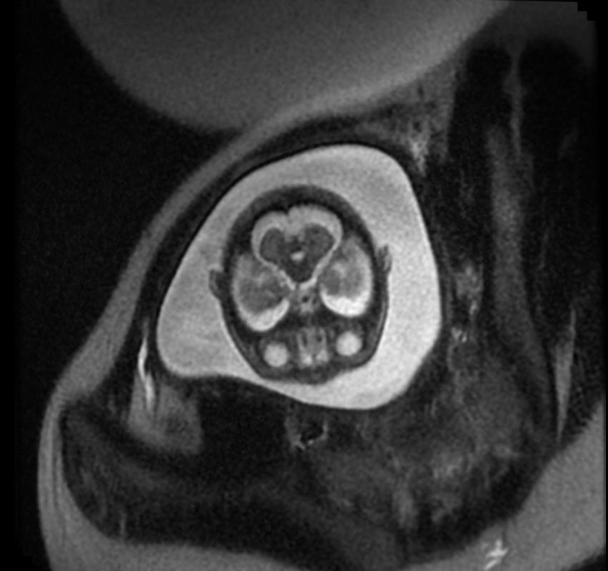
Fetal MRI: Aqueductal stenosis


Diffusion MRI
It gives better visualisation of microstructral changes in periventricular white matter areas than conventional MRI[1]