Hydrocephalus CT: Difference between revisions
No edit summary |
Kiran Singh (talk | contribs) |
||
| Line 35: | Line 35: | ||
[[Category:Neurological disorders]] | [[Category:Neurological disorders]] | ||
[[Category:Greek loanwords]] | [[Category:Greek loanwords]] | ||
[[Category:Disease]] | [[Category:Disease]] | ||
[[Category:Neurology]] | [[Category:Neurology]] | ||
Revision as of 17:18, 8 June 2015
|
Hydrocephalus Microchapters |
|
Diagnosis |
|---|
|
Treatment |
|
Case Studies |
|
Hydrocephalus CT On the Web |
|
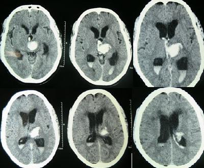
American Roentgen Ray Society Images of Hydrocephalus CT |
Editor-In-Chief: C. Michael Gibson, M.S., M.D. [1];Associate Editor-In-Chief: Cafer Zorkun M.D., PhD.,Kalsang Dolma, M.B.B.S.[2]
Overview
CT and MRI are used to evaluate ventricular size. MRI affords better imaging of the posterior fossa so can be used to evaluate posterior fossa lesion.
CT
Because the problem resides inside the head, doctors rely heavily upon computer tomography scanning (CT scans), which may be used frequently to evaluate the condition of the disorder throughout the patient's life. Each CT scan exposes the patient to many times the level of x-ray radiation of a chest x-ray. See CT radiation exposure.


Hydrocephalus
-
Hydrocephalus
-
Intracerebral hemorrhage
-
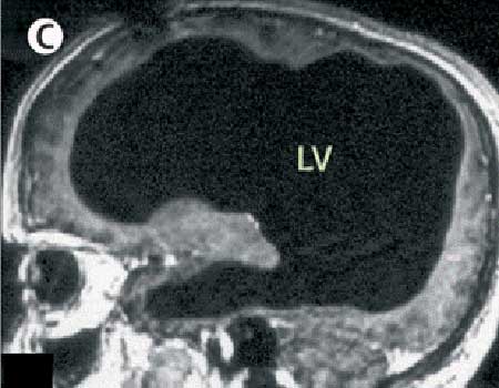
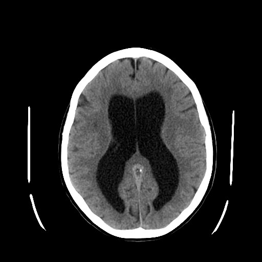
DWS: All of the black in the middle is cerebrospinal fluid and the brain matter is the rim of white along the outside of the skull.


