Hepatic hemangioma: Difference between revisions
No edit summary |
|||
| Line 1: | Line 1: | ||
__NOTOC__ | |||
{{Infobox_Disease | | {{Infobox_Disease | | ||
Name = {{PAGENAME}} | | Name = {{PAGENAME}} | | ||
| Line 9: | Line 10: | ||
OMIM = | | OMIM = | | ||
MedlinePlus = | | MedlinePlus = | | ||
MeshID = | | MeshID = | | ||
}} | }} | ||
{{ | {{Hepatic hemangioma}} | ||
'''For patient information, click [[Hepatic hemangioma (patient information)|here]]''' | '''For patient information, click [[Hepatic hemangioma (patient information)|here]]''' | ||
Revision as of 13:41, 27 November 2012
| Hepatic hemangioma | |
 | |
|---|---|
| Heptatic hemangioma. Image courtesy of RadsWiki |
|
Hepatic hemangioma Microchapters |
|
Diagnosis |
|---|
|
Treatment |
|
Case Studies |
|
Hepatic hemangioma On the Web |
|
American Roentgen Ray Society Images of Hepatic hemangioma |
For patient information, click here
Editor-In-Chief: C. Michael Gibson, M.S., M.D. [1]; Associate Editor(s)-in-Chief: Cafer Zorkun M.D., PhD.
Overview
A hepatic hemangioma is a noncancerous liver tumor made of widened (dilated) blood vessels and is the most common primary liver tumor.
Pathophysiology
- Arise from the endothelial cells that line the blood vessels and consists of multiple, large vascular channels lined by a single layer of endothelial cells and supported by collagenous walls.
- They may be associated with focal nodular hyperplasia.
- May be associated with Kasabach-Merritt syndrome: Hemolytic anemia and consumptive coagulopathy
Epidemiology and Demographics
Incidence
The incidence ranges from 0.4-20% of the population.
Age
Hepatic hemangiomas can occur at any time, but are most common in people in their 30s - 50s.
Gender
Women are affected more often than men (5:1), and usually have bigger tumors than men.
Natural History, Complications, Prognosis
In rare cases, a cavernous hemangioma may rupture. Babies may develop a type of hepatic hemangioma called benign infantile hemangioendothelioma (also called multinodular hepatic hemangiomatosis). This rare, noncancerous tumor has been linked to high rates of heart failure and death in infants. Infants are usually diagnosed by the time they are 6 months old.
Pregnancy and estrogen-based medications can cause cavernous hemangiomas to grow.
Diagnosis
They are frequently asymptomatic and incidentally discovered at imaging, surgery, or autopsy. Hepatic hemangioma is usually not discovered until medical pictures are taken of the liver for some other reason. If a cavernous hemangioma ruptures, the only sign may be an enlarged liver.
Physical Examination
Babies with benign infantile hemangioendothelioma may have:
Cardiac
- Signs of heart failure
Abdomen
Laboratory Findings
Ultrasonography
- Echogenic
- Homogenous
Computed Tomography
- Noncontrast: Hypointense to liver
- Portal venous enhancement: Peripheral nodular enhancement
- Delayed enhancement: Lesion fills in the contrast
MRI
- T2 hyperintense
- Portal venous enhancement: Peripheral nodular enhancement
- Delayed enhancement: Lesion fills in the contrast
-

T2 FrFSE
-
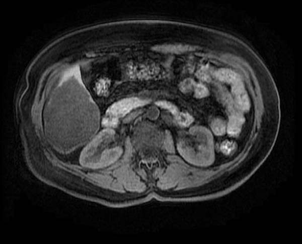
T1
-

T1 post Gad
-

T1 post Gad
-

T1 post Gad
-
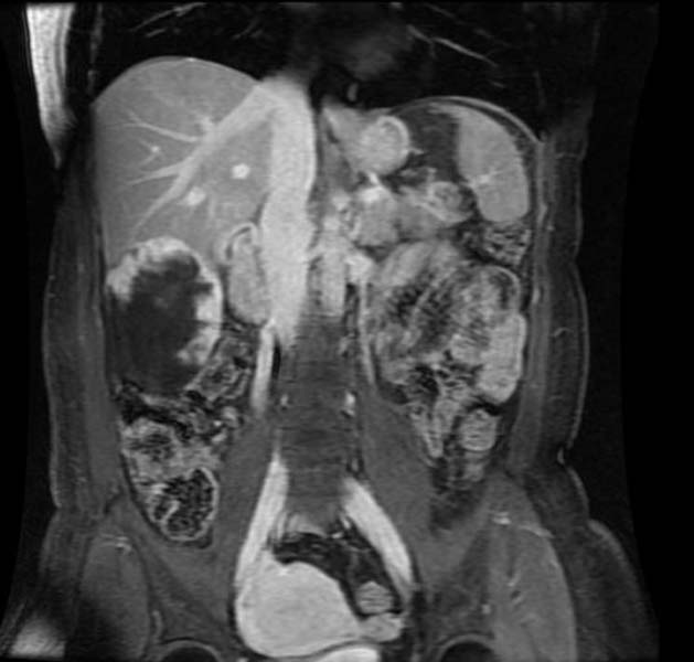
T1 post Gad coronal


Scintigraphy
- Tc99-labeled red blood cells show decreased activity on early dynamic images and increased activity on delayed images (i.e. 1-2 hours).
- Only sensitive for larger lesions.
Treatment
Most cavernous hepatic hemangiomas are treated only if there is persistent pain.
Treatment for infantile hemangioendothelioma depends on the child's growth and development. The following treatments may be needed:
- Inserting a material in a blood vessel of the liver to block it (embolization)
- Tying off (ligation) a liver artery
- Medications for heart failure
- Surgery to remove the tumor
Related Chapters
References & Additional Resources
- Moser C, Hany A, Spiegel R. [Familial giant hemangiomas of the liver. Study of a family and review of the literature]. Praxis (Bern 1994). Apr 1 1998;87(14):461-8.
- Takahashi T, Kuwao S, Katagiri H, et al. Multiple liver hemangiomas enlargement during long-term steroid therapy for myasthenia gravis. Dig Dis Sci. Jul 1998;43(7):1553-61.
- Giannitrapani L, Soresi M, La Spada E, et al. Sex hormones and risk of liver tumor. Ann N Y Acad Sci. Nov 2006;1089:228-36.
- Glinkova V, Shevah O, Boaz M, et al. Hepatic haemangiomas: possible association with female sex hormones. Gut. Sep 2004;53(9):1352-5.
- Spitzer D, Krainz R, Graf AH, et al. Pregnancy after ovarian stimulation and intrauterine insemination in a woman with cavernous macrohemangioma of the liver. A case report. J Reprod Med. Dec 1997;42(12):809-12.
- Dreyfus M, Baldauf JJ, Dadoun K, et al. Prenatal diagnosis of hepatic hemangioma. Fetal Diagn Ther. Jan-Feb 1996;11(1):57-60.
- Suzuki T, Tsuchiya N, Ito K. Multiple cavernous hemangiomas of the liver in patients with systemic lupus erythematosus. J Rheumatol. Apr 1997;24(4):810-1.
- Goodman Z. Benign tumors of the liver. In: Okuda K, Ishak KG. Neoplasms of the liver. Tokyo: Springer-Verlag; 1987:105-125.
- Mikami T, Hirata K, Oikawa I, et al. Hemobilia caused by a giant benign hemangioma of the liver: report of a case. Surg Today. 1998;28(9):948-52.
- Lee CW, Chung YH, Lee GC, et al. A case of giant hemangioma of the liver presenting with fever of unknown origin. J Korean Med Sci. Apr 1994;9(2):200-4.
- Pol B, Disdier P, Le Treut YP, et al. Inflammatory process complicating giant hemangioma of the liver: report of three cases. Liver Transpl Surg. May 1998;4(3):204-7.
- Huang SA, Tu HM, Harney JW, et al. Severe hypothyroidism caused by type 3 iodothyronine deiodinase in infantile hemangiomas. N Engl J Med. Jul 20 2000;343(3):185-9.
- Lorette G, Georgesco G, Sirinelli D, et al. [Cutaneous immature hemangioma and hepatic angioma: there is no frequent association]. Ann Dermatol Venereol. 1996;123(12):789-90.
- Brannigan M, Burns PN, Wilson SR. Blood flow patterns in focal liver lesions at microbubble-enhanced US. Radiographics. Jul-Aug 2004;24(4):921-35.
- Dietrich CF, Mertens JC, Braden B, et al. Contrast-enhanced ultrasound of histologically proven liver hemangiomas. Hepatology. May 2007;45(5):1139-45.
- von Herbay A, Vogt C, Willers R, et al. Real-time imaging with the sonographic contrast agent SonoVue: differentiation between benign and malignant hepatic lesions. J Ultrasound Med. Dec 2004;23(12):1557-68.
- Farges O, Daradkeh S, Bismuth H. Cavernous hemangiomas of the liver: are there any indications for resection?. World J Surg. Jan-Feb 1995;19(1):19-24.
- Obata S, Matsunaga N, Hayashi K, et al. Fluid-fluid levels in giant cavernous hemangioma of the liver: CT and MRI demonstration. Abdom Imaging. Nov-Dec 1998;23(6):600-2.
- Kinnard MF, Alavi A, Rubin RA, et al. Nuclear imaging of solid hepatic masses. Semin Roentgenol. Oct 1995;30(4):375-95.
- Krause T, Hauenstein K, Studier-Fischer B, et al. Improved evaluation of technetium-99m-red blood cell SPECT in hemangioma of the liver. J Nucl Med. Mar 1993;34(3):375-80.
- Tsai CC, Yen TC, Tzen KY. Pedunculated giant liver hemangioma mimicking a hypervascular gastric tumor on Tc-99m RBC SPECT. Clin Nucl Med. Feb 1999;24(2):132-3.
- De Franco A, Monteforte MG, Maresca G, et al. [Integrated diagnosis of liver angioma: comparison of Doppler color ultrasonography, computerized tomography, and magnetic resonance]. Radiol Med. Jan-Feb 1997;93(1-2):87-94.
- Heilo A, Stenwig AE. Liver hemangioma: US-guided 18-gauge core-needle biopsy. Radiology. Sep 1997;204(3):719-22.
- Bruix J, Sherman M, Llovet JM, et al. Clinical management of hepatocellular carcinoma. Conclusions of the Barcelona-2000 EASL conference. European Association for the Study of the Liver. J Hepatol. Sep 2001;35(3):421-30.
- Craig JR, Peters RL, Edmondson HA. Tumors of the Liver and Intrahepatic Bile Ducts, 2nd series, fascicle 26. Washington, DC. Armed Forces Institute of Pathology. 1989;191.
- Ishak KG, Markin RS. Liver. In: Damjanov I, Linder J, eds. Anderson's Pathology. 10th ed. Mosby: St. Louis, Mo; 1996:1834.
- Gibney RG, Hendin AP, Cooperberg PL. Sonographically detected hepatic hemangiomas: absence of change over time. AJR Am J Roentgenol. Nov 1987;149(5):953-7.
- Corigliano N, Mercantini P, Amodio PM, et al. Hemoperitoneum from a spontaneous rupture of a giant hemangioma of the liver: report of a case. Surg Today. 2003;33(6):459-63.
- Arnoletti JP, Brodsky J. Surgical treatment of benign hepatic mass lesions. Am Surg. May 1999;65(5):431-3.
- Srivastava DN, Gandhi D, Seith A, et al. Transcatheter arterial embolization in the treatment of symptomatic cavernous hemangiomas of the liver: a prospective study. Abdom Imaging. Sep-Oct 2001;26(5):510-4.
- Zeng Q, Li Y, Chen Y, et al. Gigantic cavernous hemangioma of the liver treated by intra-arterial embolization with pingyangmycin-lipiodol emulsion: a multi-center study. Cardiovasc Intervent Radiol. Sep-Oct 2004;27(5):481-5.
- Rokitansky AM, Jakl RJ, Gopfrich H, et al. Special compression sutures: a new surgical technique to achieve a quick decrease in shunt volume caused by diffuse hemangiomatosis of the liver. Pediatr Surg Int. Nov 1998;14(1-2):119-21.
- Tak WY, Park SY, Jeon SW, et al. Ultrasonography-guided percutaneous radiofrequency ablation for treatment of a huge symptomatic hepatic cavernous hemangioma. J Clin Gastroenterol. Feb 2006;40(2):167-70.
- Fan RF, Chai FL, He GX, et al. Laparoscopic radiofrequency ablation of hepatic cavernous hemangioma. A preliminary experience with 27 patients. Surg Endosc. Feb 2006;20(2):281-5.
- Biswal BM, Sandhu M, Lal P, et al. Role of radiotherapy in cavernous hemangioma liver. Indian J Gastroenterol. Jul 1995;14(3):95-8.
- Tepetes K, Selby R, Webb M, et al. Orthotopic liver transplantation for benign hepatic neoplasms. Arch Surg. Feb 1995;130(2):153-6.