Gastrointestinal stromal tumor other imaging findings: Difference between revisions
Akshun Kalia (talk | contribs) |
|||
| (4 intermediate revisions by 2 users not shown) | |||
| Line 7: | Line 7: | ||
==Other Imaging Findings== | ==Other Imaging Findings== | ||
[[Endoscopy]] may be helpful in the diagnosis of gastrointestinal stromal tumor (GIST). An [[endoscope]] can be used in conditions where GIST is located in accessible places such as stomach, esophagus and large intestine. However, GIST located outside the [[lumen]] of wall may not be visible on an [[endoscopy]]. Findings on an [[endoscopy]] suggestive of GIST include:<ref name="pmid11747229">{{cite journal |vauthors=Gu M, Ghafari S, Nguyen PT, Lin F |title=Cytologic diagnosis of gastrointestinal stromal tumors of the stomach by endoscopic ultrasound-guided fine-needle aspiration biopsy: cytomorphologic and immunohistochemical study of 12 cases |journal=Diagn. Cytopathol. |volume=25 |issue=6 |pages=343–50 |year=2001 |pmid=11747229 |doi= |url=}}</ref><ref name="pmid12376922">{{cite journal |vauthors=Fu K, Eloubeidi MA, Jhala NC, Jhala D, Chhieng DC, Eltoum IE |title=Diagnosis of gastrointestinal stromal tumor by endoscopic ultrasound-guided fine needle aspiration biopsy--a potential pitfall |journal=Ann Diagn Pathol |volume=6 |issue=5 |pages=294–301 |year=2002 |pmid=12376922 |doi= |url=}}</ref><ref name="pmid22943011">{{cite journal |vauthors=Zhao X, Yue C |title=Gastrointestinal stromal tumor |journal=J Gastrointest Oncol |volume=3 |issue=3 |pages=189–208 |year=2012 |pmid=22943011 |pmc=3418531 |doi=10.3978/j.issn.2078-6891.2012.031 |url=}}</ref> | [[Endoscopy]] may be helpful in the diagnosis of gastrointestinal stromal tumor (GIST). An [[endoscope]] can be used in conditions where GIST is located in accessible places such as [[stomach]], [[esophagus]] and [[Intestine|large intestine.]] However, GIST located outside the [[lumen]] of wall may not be visible on an [[endoscopy]]. Findings on an [[endoscopy]] suggestive of GIST include:<ref name="pmid11747229">{{cite journal |vauthors=Gu M, Ghafari S, Nguyen PT, Lin F |title=Cytologic diagnosis of gastrointestinal stromal tumors of the stomach by endoscopic ultrasound-guided fine-needle aspiration biopsy: cytomorphologic and immunohistochemical study of 12 cases |journal=Diagn. Cytopathol. |volume=25 |issue=6 |pages=343–50 |year=2001 |pmid=11747229 |doi= |url=}}</ref><ref name="pmid12376922">{{cite journal |vauthors=Fu K, Eloubeidi MA, Jhala NC, Jhala D, Chhieng DC, Eltoum IE |title=Diagnosis of gastrointestinal stromal tumor by endoscopic ultrasound-guided fine needle aspiration biopsy--a potential pitfall |journal=Ann Diagn Pathol |volume=6 |issue=5 |pages=294–301 |year=2002 |pmid=12376922 |doi= |url=}}</ref><ref name="pmid22943011">{{cite journal |vauthors=Zhao X, Yue C |title=Gastrointestinal stromal tumor |journal=J Gastrointest Oncol |volume=3 |issue=3 |pages=189–208 |year=2012 |pmid=22943011 |pmc=3418531 |doi=10.3978/j.issn.2078-6891.2012.031 |url=}}</ref> | ||
*Smooth submucosal mass | *Smooth [[submucosal]] [[mass]] | ||
*Areas of [[ulceration]] or [[bleeding]] | *Areas of [[ulceration]] or [[bleeding]] | ||
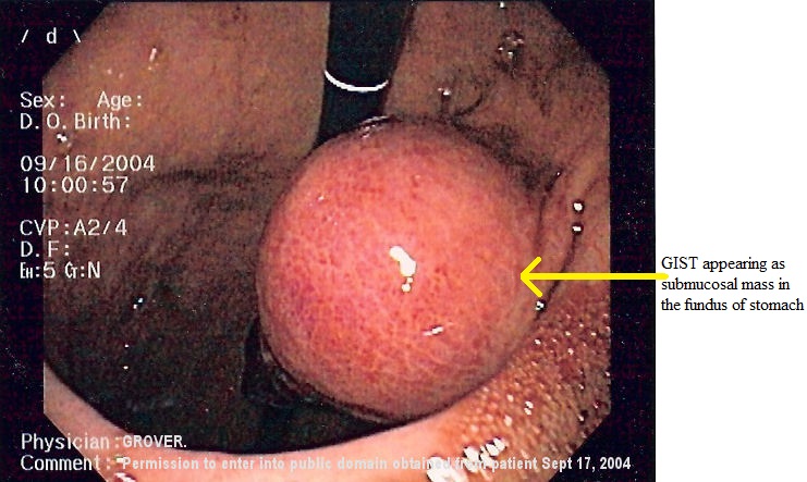
[[Image:GIST 2.jpg|thumb|left|[[Endoscopy|Endoscopic]] image of GIST in the fundus of [[stomach]]. ([Courtesy: By Samir, CC BY-SA 3.0, https://commons.wikimedia.org/w/index.php?curid=1257100])]] | [[Image:GIST 2.jpg|thumb|left|[[Endoscopy|Endoscopic]] image of GIST in the fundus of [[stomach]]. ([Courtesy: By Samir, CC BY-SA 3.0, https://commons.wikimedia.org/w/index.php?curid=1257100])]] | ||
{| | |||
|- | |||
| | |||
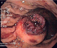
[[image:GIST 3.jpg|thumb|center|[[Endoscopy|Endoscopic]] image of GIST in the fundus of stomach with an overlying clot. ([Courtesy: By Samir (http://en.wikipedia.org/wiki/Image:GIST_3.jpg) [GFDL (http://www.gnu.org/copyleft/fdl.html) or CC-BY-SA-3.0 (http://creativecommons.org/licenses/by-sa/3.0/)], via Wikimedia Commons])]] | [[image:GIST 3.jpg|thumb|center|[[Endoscopy|Endoscopic]] image of GIST in the fundus of stomach with an overlying clot. ([Courtesy: By Samir (http://en.wikipedia.org/wiki/Image:GIST_3.jpg) [GFDL (http://www.gnu.org/copyleft/fdl.html) or CC-BY-SA-3.0 (http://creativecommons.org/licenses/by-sa/3.0/)], via Wikimedia Commons])]] | ||
Endoscopic guided biopsy may be done for definite diagnosis of gastrointestinal stromal tumor (GIST). However, percutaneous biopsy is not routinely recommended. | |- | ||
*Patients with unresectable GIST must undergo biopsy to determine tumor cell type and chemotherapy. | |} | ||
*GISTs are highly vascular which puts them at a risk of bleeding. | [[Endoscopy|Endoscopic]] guided biopsy may be done for definite diagnosis of gastrointestinal stromal tumor (GIST). However, percutaneous [[biopsy]] is not routinely recommended. | ||
**Percutaneous fine needle biopsy may put the patient at an increased risk of tumor rupture and bleeding. | *[[Patient|Patients]] with unresectable GIST must undergo [[biopsy]] to determine [[tumor]] [[Cell (biology)|cell]] type and [[chemotherapy]]. | ||
**Percutaneous biopsy can also lead to tumor seeding along the biopsy tract such as peritoneum or mesentery. | *GISTs are highly [[vascular]] which puts them at a risk of [[bleeding]]. | ||
**Thus, patients in whom surgery is an option are advised not to undergo biopsy. | **[[Percutaneous]] fine needle [[biopsy]] may put the [[patient]] at an increased risk of [[tumor]] rupture and [[bleeding]]. | ||
**[[Percutaneous]] [[biopsy]] can also lead to [[tumor]] seeding along the [[biopsy]] tract such as [[peritoneum]] or [[mesentery]]. | |||
**Thus, [[Patient|patients]] in whom surgery is an option are advised not to undergo [[biopsy]]. | |||
==References== | ==References== | ||
Latest revision as of 03:36, 4 March 2019
|
Gastrointestinal stromal tumor Microchapters |
|
Differentiating Gastrointestinal stromal tumor from other Diseases |
|---|
|
Diagnosis |
|
Treatment |
|
Case Studies |
|
Gastrointestinal stromal tumor other imaging findings On the Web |
|
American Roentgen Ray Society Images of Gastrointestinal stromal tumor other imaging findings |
|
FDA on Gastrointestinal stromal tumor other imaging findings |
|
CDC on Gastrointestinal stromal tumor other imaging findings |
|
Gastrointestinal stromal tumor other imaging findings in the news |
|
Blogs on Gastrointestinal stromal tumor other imaging findings |
|
Directions to Hospitals Treating Gastrointestinal stromal tumor |
|
Risk calculators and risk factors for Gastrointestinal stromal tumor other imaging findings |
Editor-In-Chief: C. Michael Gibson, M.S., M.D. [1]Associate Editor(s)-in-Chief: Akshun Kalia M.B.B.S.[2]
Overview
Endoscopy may be helpful in the diagnosis of gastrointestinal stromal tumor (GIST).An endoscope can be used in conditions where GIST is located in accessible places such as stomach, esophagus and large intestine. On an endoscopy, GIST can appear as a smooth submucosal mass with areas of ulceration or bleeding.
Other Imaging Findings
Endoscopy may be helpful in the diagnosis of gastrointestinal stromal tumor (GIST). An endoscope can be used in conditions where GIST is located in accessible places such as stomach, esophagus and large intestine. However, GIST located outside the lumen of wall may not be visible on an endoscopy. Findings on an endoscopy suggestive of GIST include:[1][2][3]
- Smooth submucosal mass
- Areas of ulceration or bleeding

 |
Endoscopic guided biopsy may be done for definite diagnosis of gastrointestinal stromal tumor (GIST). However, percutaneous biopsy is not routinely recommended.
- Patients with unresectable GIST must undergo biopsy to determine tumor cell type and chemotherapy.
- GISTs are highly vascular which puts them at a risk of bleeding.
- Percutaneous fine needle biopsy may put the patient at an increased risk of tumor rupture and bleeding.
- Percutaneous biopsy can also lead to tumor seeding along the biopsy tract such as peritoneum or mesentery.
- Thus, patients in whom surgery is an option are advised not to undergo biopsy.
References
- ↑ Gu M, Ghafari S, Nguyen PT, Lin F (2001). "Cytologic diagnosis of gastrointestinal stromal tumors of the stomach by endoscopic ultrasound-guided fine-needle aspiration biopsy: cytomorphologic and immunohistochemical study of 12 cases". Diagn. Cytopathol. 25 (6): 343–50. PMID 11747229.
- ↑ Fu K, Eloubeidi MA, Jhala NC, Jhala D, Chhieng DC, Eltoum IE (2002). "Diagnosis of gastrointestinal stromal tumor by endoscopic ultrasound-guided fine needle aspiration biopsy--a potential pitfall". Ann Diagn Pathol. 6 (5): 294–301. PMID 12376922.
- ↑ Zhao X, Yue C (2012). "Gastrointestinal stromal tumor". J Gastrointest Oncol. 3 (3): 189–208. doi:10.3978/j.issn.2078-6891.2012.031. PMC 3418531. PMID 22943011.