Gallbladder cancer echocardiography or ultrasound: Difference between revisions
Varun Kumar (talk | contribs) (Created page with "{{Gallbladder cancer}} {{CMG}} ==Overview== ==References== {{reflist|2}} Category:Disease Category:Types of cancer Category:Oncology [[Category:Gastroenterolog...") |
Varun Kumar (talk | contribs) |
||
| Line 3: | Line 3: | ||
==Overview== | ==Overview== | ||
==Ultrasonography== | |||
[http://www.peir.net Images courtesy of Professor Peter Anderson DVM PhD and published with permission © PEIR, University of Alabama at Birmingham, Department of Pathology] | |||
<div align="left"> | |||
<gallery heights="150" widths="150"> | |||
Image:Gallbladder cancer usg 1.jpg|Gallbladder cancer: 88 year old female with epigastric pain, nausea and vomiting. Soft tissue mass lesion arising from the anterior wall of the gallbladder and extending into the gallbladder lumen. | |||
Image:Gallbladder cancer usg 2.jpg|Gallbladder cancer: 88 year old female with epigastric pain, nausea and vomiting. This has the appearance of a neoplasm, most likely a primary carcinoma of the gallbladder. | |||
</gallery> | |||
</div> | |||
<div align="left"> | |||
<gallery heights="150" widths="150"> | |||
Image:Gallbladder cancer usg 3.jpg|Gallbladder cancer: 88 year old female with epigastric pain, nausea and vomiting. Metastatic lesion or lymphoma are less likely considerations. There is no definite sonographic evidence of extension of the mass outside the gallbladder nor of regional adenopathy. | |||
Image:Gallbladder cancer usg 4.jpg|Gallbladder cancer: 88 year old female with epigastric pain, nausea and vomiting. Bilateral renal cysts, with that in the left upper pole categorized as a Bosniak type II cyst. | |||
</gallery> | |||
</div> | |||
==References== | ==References== | ||
Revision as of 02:21, 22 January 2012
|
Gallbladder cancer Microchapters |
|
Diagnosis |
|---|
|
Treatment |
|
Case Studies |
|
Gallbladder cancer echocardiography or ultrasound On the Web |
|
American Roentgen Ray Society Images of Gallbladder cancer echocardiography or ultrasound |
|
Gallbladder cancer echocardiography or ultrasound in the news |
|
Risk calculators and risk factors for Gallbladder cancer echocardiography or ultrasound |
Editor-In-Chief: C. Michael Gibson, M.S., M.D. [1]
Overview
Ultrasonography
-
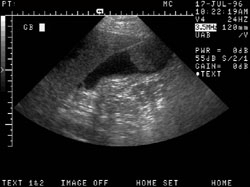
Gallbladder cancer: 88 year old female with epigastric pain, nausea and vomiting. Soft tissue mass lesion arising from the anterior wall of the gallbladder and extending into the gallbladder lumen.
-
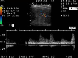
Gallbladder cancer: 88 year old female with epigastric pain, nausea and vomiting. This has the appearance of a neoplasm, most likely a primary carcinoma of the gallbladder.


-
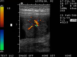
Gallbladder cancer: 88 year old female with epigastric pain, nausea and vomiting. Metastatic lesion or lymphoma are less likely considerations. There is no definite sonographic evidence of extension of the mass outside the gallbladder nor of regional adenopathy.
-
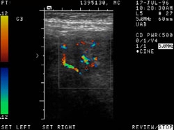
Gallbladder cancer: 88 year old female with epigastric pain, nausea and vomiting. Bilateral renal cysts, with that in the left upper pole categorized as a Bosniak type II cyst.


References