Echinococcosis x ray: Difference between revisions
Jump to navigation
Jump to search
Aditya Ganti (talk | contribs) |
m (Bot: Removing from Primary care) |
||
| (9 intermediate revisions by 5 users not shown) | |||
| Line 1: | Line 1: | ||
__NOTOC__ | __NOTOC__ | ||
{{Echinococcosis}} | {{Echinococcosis}} | ||
{{CMG}} '''Associate Editor-In-Chief:''' {{CZ}}; {{KD}} | {{CMG}} '''Associate Editor-In-Chief:''' {{MIR}} ; {{CZ}}; {{KD}} | ||
==Overview== | ==Overview== | ||
Radiography imaging permits the detection of hydatid cysts in the lungs; however, in other organ sites, calcifications can be visualized. On a chest | [[Radiography]] [[imaging]] permits the detection of [[Hydatid cyst|hydatid cysts]] in the [[Lung|lungs]]; however, in other [[Organ (anatomy)|organ]] sites, [[Calcification|calcifications]] can be visualized. On a chest x-ray, [[cysts]] are well defined as a rounded mass with uniform [[density]].<ref name="pmid18784219">{{cite journal |vauthors=Junghanss T, da Silva AM, Horton J, Chiodini PL, Brunetti E |title=Clinical management of cystic echinococcosis: state of the art, problems, and perspectives |journal=Am. J. Trop. Med. Hyg. |volume=79 |issue=3 |pages=301–11 |year=2008 |pmid=18784219 |doi= |url=}}</ref> | ||
==X-ray== | ==X-ray== | ||
Radiography permits the detection of hydatid cysts in the lungs; however, in other organ sites, | [[Radiography]] permits the detection of [[Hydatid cyst|hydatid cysts]] in the [[lungs]]; however, in other [[Organ (anatomy)|organ]] sites, [[Calcification|calcifications]] if present can be visualized. <ref name="pmid18784219">{{cite journal |vauthors=Junghanss T, da Silva AM, Horton J, Chiodini PL, Brunetti E |title=Clinical management of cystic echinococcosis: state of the art, problems, and perspectives |journal=Am. J. Trop. Med. Hyg. |volume=79 |issue=3 |pages=301–11 |year=2008 |pmid=18784219 |doi= |url=}}</ref> | ||
*On a [[Chest X-ray|chest x-ray]], [[Cyst|cysts]] are well defined as a rounded [[mass]] with uniform density. | |||
*If [[cyst]] ruptures endocyst gets detached from the [[membranes]] of the [[cyst]] and is seen floating within the [[cyst]]. ("water-lily" or "meniscus" sign) | |||
[[File:Webp.net-gifmaker (38).gif|500px|center|thumb|<small><small>Courtesy dedicated to radiopaedia.com</small></small>]] | |||
== References == | == References == | ||
{{reflist|2}} | {{reflist|2}} | ||
{{WH}} | |||
{{WS}} | |||
[[Category:Parasitic diseases]] | [[Category:Parasitic diseases]] | ||
[[Category:Disease]] | [[Category:Disease]] | ||
[[Category:Needs content]] | [[Category:Needs content]] | ||
[[Category:Emergency medicine]] | |||
[[Category:Up-To-Date]] | |||
[[Category:Infectious disease]] | |||
[[Category:Hepatology]] | |||
[[Category:Gastroenterology]] | |||
[[Category:Surgery]] | |||
Latest revision as of 21:32, 29 July 2020
|
Echinococcosis Microchapters |
|
Diagnosis |
|---|
|
Treatment |
|
Case Studies |
|
Echinococcosis x ray On the Web |
|
American Roentgen Ray Society Images of Echinococcosis x ray |
Editor-In-Chief: C. Michael Gibson, M.S., M.D. [1] Associate Editor-In-Chief: Mahshid Mir, M.D. [2] ; Cafer Zorkun, M.D., Ph.D. [3]; Kalsang Dolma, M.B.B.S.[4]
Overview
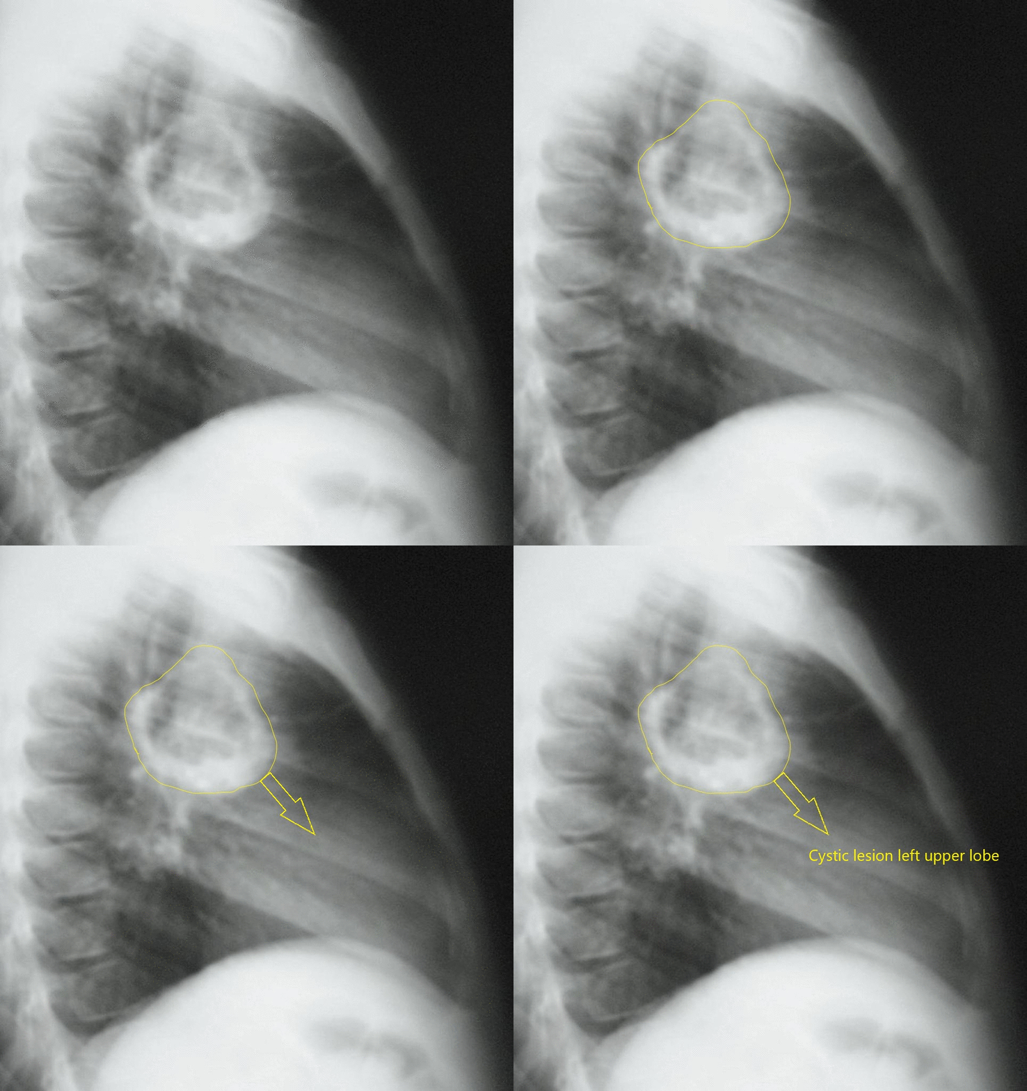
Radiography imaging permits the detection of hydatid cysts in the lungs; however, in other organ sites, calcifications can be visualized. On a chest x-ray, cysts are well defined as a rounded mass with uniform density.[1]
X-ray
Radiography permits the detection of hydatid cysts in the lungs; however, in other organ sites, calcifications if present can be visualized. [1]
- On a chest x-ray, cysts are well defined as a rounded mass with uniform density.
- If cyst ruptures endocyst gets detached from the membranes of the cyst and is seen floating within the cyst. ("water-lily" or "meniscus" sign)
