Conjunctivitis physical examination: Difference between revisions
No edit summary |
No edit summary |
||
| Line 12: | Line 12: | ||
*[[Hyperemia]] | *[[Hyperemia]] | ||
*[[Chemosis]] | *[[Chemosis]] | ||
* | *Follicular conjunctival reaction | ||
*Pseudomembranous (occasionally) | *Pseudomembranous (occasionally) | ||
*[[Cicatricial]] [[conjunctival]] reaction | *[[Cicatricial]] [[conjunctival]] reaction | ||
| Line 19: | Line 19: | ||
===Acute Hemorrhagic Conjunctivitis=== | ===Acute Hemorrhagic Conjunctivitis=== | ||
Eye examination of patients with [[acute]] hemorrhagic conjunctivitis is usually remarkable for: | Eye examination of patients with [[acute]] hemorrhagic conjunctivitis is usually remarkable for: | ||
*[[Swollen]], | *[[Swollen]], Edematous [[Eyelid]] | ||
*Eye pain in [[ | *Eye pain in [[palpation]] | ||
*[[Bulbar]] [[conjunctiva]] [[hemorrhage]] | *[[Bulbar]] [[conjunctiva]] [[hemorrhage]] | ||
===Bacterial | ===Bacterial Conjunctivitis=== | ||
Eye examination of patients with [[bacterial]] conjunctivitis is usually remarkable for: | Eye examination of patients with [[bacterial]] conjunctivitis is usually remarkable for: | ||
*[[Bulbar]] [[conjunctival]] injection | *[[Bulbar]] [[conjunctival]] injection | ||
* | *Palpebral [[conjunctival]] [[papillary]] reaction | ||
* | *Muco-purulent or watery discharge | ||
*[[Chemosis]] | *[[Chemosis]] | ||
*Lid [[erythema]] | *Lid [[erythema]] | ||
===Neonatal Conjunctivitis=== | |||
Eye examination of patients with [[neonatal]] conjunctivitis is usually remarkable for: | |||
*''[[Neisseria gonorrhea]]'' | |||
**Chemosis | |||
**[[Severe]] [[lid edema]] and | |||
**[[Mucopurulent]] discharge | |||
**[[Corneal]] involvement (diffuse [[epithelial edema]], [[ulceration]], corneal [[perforation]], and [[endophthalmitis]] | |||
*''[[Chlamydia trachomatis]]'' | |||
**Mild [[hyperemia]] | |||
**Mucoid discharge | |||
**[[Chemosis]] | |||
**Pseudomembrane formation | |||
*Chemical | |||
**Mild conjunctival [[injection]] | |||
**Tearing | |||
[[Redness]] of the [[conjunctiva]] on one or both eyes should be apparent, but may be quite mild. Except in obvious pyogenic or toxic/chemical conjunctivitis, a [[slit lamp]] (biomicroscope) is needed to have any confidence in the diagnosis. Examination of the tarsal conjunctiva is usually more diagnostic than the bulbar conjunctiva. | [[Redness]] of the [[conjunctiva]] on one or both eyes should be apparent, but may be quite mild. Except in obvious pyogenic or toxic/chemical conjunctivitis, a [[slit lamp]] (biomicroscope) is needed to have any confidence in the diagnosis. Examination of the tarsal conjunctiva is usually more diagnostic than the bulbar conjunctiva. | ||
Revision as of 17:08, 1 July 2016
|
Conjunctivitis Microchapters |
|
Diagnosis |
|---|
|
Treatment |
|
Case Studies |
|
Conjunctivitis physical examination On the Web |
|
American Roentgen Ray Society Images of Conjunctivitis physical examination |
|
Risk calculators and risk factors for Conjunctivitis physical examination |
Editor-In-Chief: C. Michael Gibson, M.S., M.D. [1] Associate Editor(s)-in-Chief: Sara Mehrsefat, M.D. [2]
Overview
Infection (redness) of the conjunctiva on one or both eyes should be apparent, but may be quite mild. Except in obvious pyogenic or toxic/chemical conjunctivitis, a slit lamp (biomicroscope) is needed to have any confidence in the diagnosis. Examination of the tarsal conjunctiva is usually more diagnostic than the bulbar conjunctiva.
Physical Examination
Viral Conjunctivitis
Patients with viral conjunctivitis usually appear febrile, and they have Preauricular adenopathy. Eye examination of patients with viral conjunctivitis is usually remarkable for:
- Epiphora
- Hyperemia
- Chemosis
- Follicular conjunctival reaction
- Pseudomembranous (occasionally)
- Cicatricial conjunctival reaction
- Edematous and ecchymotic eyelids
- Corneal epithelial defect (severe cases)
Acute Hemorrhagic Conjunctivitis
Eye examination of patients with acute hemorrhagic conjunctivitis is usually remarkable for:
- Swollen, Edematous Eyelid
- Eye pain in palpation
- Bulbar conjunctiva hemorrhage
Bacterial Conjunctivitis
Eye examination of patients with bacterial conjunctivitis is usually remarkable for:
- Bulbar conjunctival injection
- Palpebral conjunctival papillary reaction
- Muco-purulent or watery discharge
- Chemosis
- Lid erythema
Neonatal Conjunctivitis
Eye examination of patients with neonatal conjunctivitis is usually remarkable for:
- Neisseria gonorrhea
- Chemosis
- Severe lid edema and
- Mucopurulent discharge
- Corneal involvement (diffuse epithelial edema, ulceration, corneal perforation, and endophthalmitis
- Chlamydia trachomatis
- Chemical
- Mild conjunctival injection
- Tearing
Redness of the conjunctiva on one or both eyes should be apparent, but may be quite mild. Except in obvious pyogenic or toxic/chemical conjunctivitis, a slit lamp (biomicroscope) is needed to have any confidence in the diagnosis. Examination of the tarsal conjunctiva is usually more diagnostic than the bulbar conjunctiva.
Allergic conjunctivitisshows pale watery swelling or edema of the conjunctiva and sometimes the whole eyelid, often with a ropy, non-purulent mucoid discharge. There is variable redness. Viral conjunctivitis, commonly known as "pink eye", shows a fine diffuse pinkness of the conjunctiva which is easily mistaken for the 'ciliary infection' of iritis, but there are usually corroborative signs on biomicroscopy, particularly numerous lymphoid follicle12:02, 22 January 2008 (EST)12:02, 22 January 2008 (EST)~~s on the tarsal conjunctiva, and sometimes a punctate keratitis.
Pyogenic bacterial conjunctivitis shows an opaque purulent discharge, a very red eye, and on biomicroscopy there are numerous white cells and desquamated epithelial cells seen in the 'tear gutter' along the lid margin. The tarsal conjunctiva is a velvety red and not particularly follicular. Non-pyogenic infections can show just mild injection and be difficult to diagnose. Scarring of the tarsal conjunctiva is occasionally seen in chronic infections, especially in trachoma.
Irritant or toxic conjunctivitis show primarily marked redness. If due to splash injury, it is often present only in the lower conjunctival sac. With some chemicals—above all with caustic alkalis such as sodium hydroxide—there may be necrosis of the conjunctiva with a deceptively white eye due to vascular closure, followed by sloughing of the dead epithelium. This is likely to be associated with slit-lamp evidence of anterior uveitis.
Images
The following are gross images associated with rheumatic fever.[1]
-
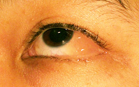
One eye with conjunctivitis.
-
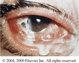
Bacterial Conjunctivitis
-
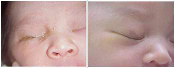
Chemical Conjunctivitis


.jpg)
(Images shown below are courtesy of Charlie Goldberg, M.D., UCSD School of Medicine and VA Medical Center, San Diego, CA)
-
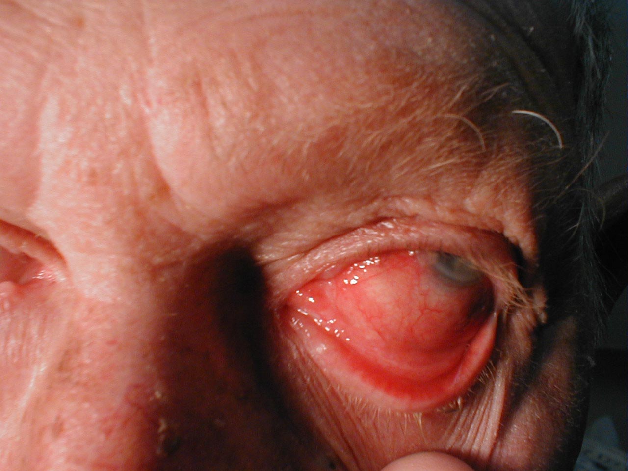
Conjunctivitis: Note inflamed conjunctiva of sclera and reflection onto underside of eyelid.
-
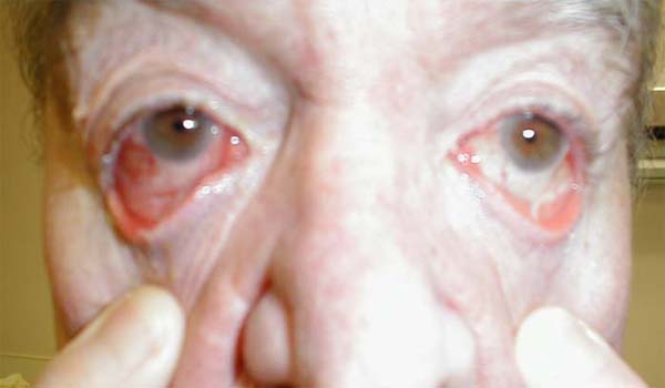
Conjunctivitis: Marked bilateral inflammation involving conjunctiva that covers sclera and under surface of eyelid. Thick exudate can also be seen.
-
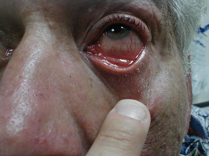
Conjunctivitis: Inflammation of conjunctiva covering sclera and under surface of eyelid.


