Bursitis classification: Difference between revisions
| Line 12: | Line 12: | ||
Common anatomic location include: | Common anatomic location include: | ||
*Elbow | *Elbow | ||
**Olecranon bursa | **[[Olecranon bursitis|[Olecranon bursa]] | ||
*Shoulder | *Shoulder | ||
**Subacromial-subdeltoid bursa (most common) | **[[Subacromial bursitis|Subacromial-subdeltoid bursa]] (most common) | ||
**Subscapular recess | **Subscapular recess | ||
**Subcoracoid bursa | **Subcoracoid bursa | ||
| Line 20: | Line 20: | ||
**Supra-acromial bursa | **Supra-acromial bursa | ||
*Hip | *Hip | ||
**[[Trochanteric bursitis|Trochanteric bursa]] | |||
**Iliopsoas bursa | **Iliopsoas bursa | ||
**Ischal bursa | **Ischal bursa | ||
*Knee | *Knee | ||
**[[Prepatellar bursitis|Prepatellar bursa]] | |||
**Infrapatellar bursa | **Infrapatellar bursa | ||
**Suprapatellar bursa | **Suprapatellar bursa | ||
*Ankle | *Ankle | ||
**Retrocalcaneal bursa | **[[Retrocalcaneal bursitis|Retrocalcaneal bursa]] | ||
**Achilles bursea | **Achilles bursea | ||
Based on the location of the bursa from the skin, bursitis may be classified into 2 subtyopes: superficial and deep | Based on the location of the bursa from the skin, bursitis may be classified into 2 subtyopes: superficial and deep | ||
Revision as of 13:36, 22 August 2016
|
Bursitis Microchapters |
|
Diagnosis |
|---|
|
Treatment |
|
Case Studies |
|
Bursitis classification On the Web |
|
American Roentgen Ray Society Images of Bursitis classification |
|
Risk calculators and risk factors for Bursitis classification |
Editor-In-Chief: C. Michael Gibson, M.S., M.D. [1]; Associate Editor(s)-in-Chief: Sara Mehrsefat, M.D. [2]
Overview
Classification
Based on the nature of inflammation bursitis may classified into:[1]
Common anatomic location include:
- Elbow
- Shoulder
- Subacromial-subdeltoid bursa (most common)
- Subscapular recess
- Subcoracoid bursa
- Coracoclavicular bursa
- Supra-acromial bursa
- Hip
- Trochanteric bursa
- Iliopsoas bursa
- Ischal bursa
- Knee
- Prepatellar bursa
- Infrapatellar bursa
- Suprapatellar bursa
- Ankle
- Retrocalcaneal bursa
- Achilles bursea
Based on the location of the bursa from the skin, bursitis may be classified into 2 subtyopes: superficial and deep
- Common superficial form of bursitis include:
- Olecranon bursitis
- Pre-patellar bursitis
- Infrapatellar bursitis
- Retrocalcanea bursitis
- Common deep form of bursitis include:
- Anserine
- Subacromial
- Trochanteric
Additionally, based on duration of symptoms and presentation bursitis may classified into: acute, subacute and chronic
Images
The following are images associated with different type of bursitis.[1]
-

(1)Subacromial-subdeltoid bursa (2) Subscapular recess (3) Subcoracoid bursa (4) Coracoclavicular bursa (5) Supra-acromial bursa (6) Medial extension of subacromial-subdeltoid bursa
-

Subcoracoid bursa
-

Superficial and deep infrapatellar bursae.
-

Prepatellar bursa
-

Pes anserine bursitis
-
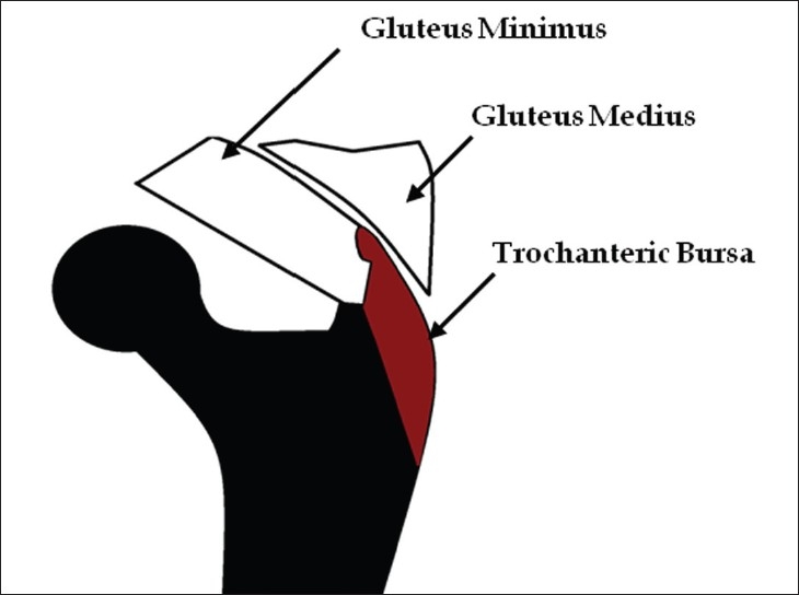
Trochanteric bursa
-
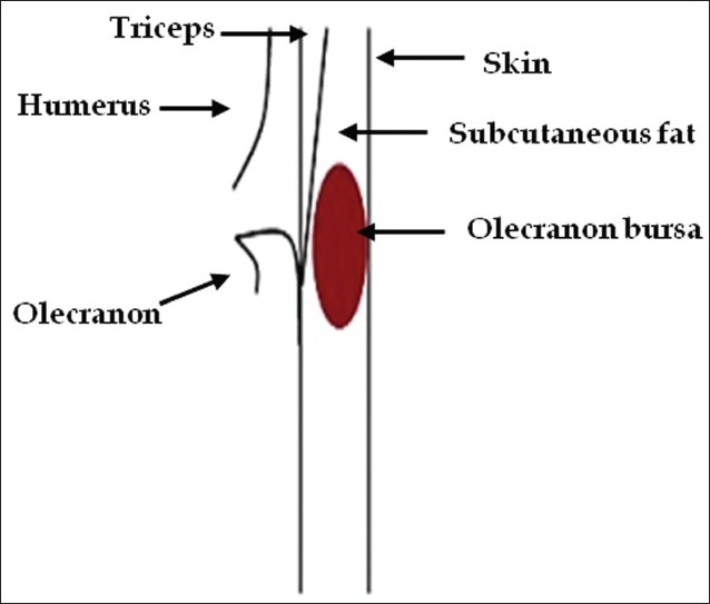
Olecranon bursa


References
- ↑ 1.0 1.1 Chatra PS (2012). "Bursae around the knee joints". Indian J Radiol Imaging. 22 (1): 27–30. doi:10.4103/0971-3026.95400. PMC 3354353. PMID 22623812.