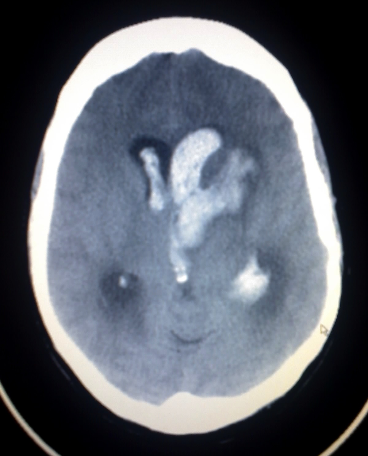
Bonnet-Dechaume-Blanc syndrome
Editor-In-Chief: C. Michael Gibson, M.S., M.D. [1] Associate Editor(s)-in-Chief: Jyostna Chouturi, M.B.B.S [2]
Synonyms and keywords: Wyburn mason's syndrome; Retinoencephalofacial angiomatosis

Overview
.
Signs and symptoms
Causes
The syndrome is a congenital disorder that begins to develop around the seventh week of gestation when the maturation of retinal mesenchymal cells do not properly grow.[1][2][3] The abnormal development of vascular tissue leads to arteriovenous malformations. These malformations affect both the visual and cerebral structures and lead to the development of the syndrome.[3]
Mechanism
Bonnet–Dechaume–Blanc syndrome results mainly from arteriovenous malformations. These malformations are addressed previously in the article, under “Signs and Symptoms.” Due to lack of research, it is difficult to provide a specific mechanism for this disorder. However, a number of examinations, mentioned under “Diagnosis,” can be performed on subjects to investigate the disorder and severity of the AVMs.
Epidemiology
The syndrome was first described in 1943 and believed to be associated with racemose hemangiomatosis of the retina and arteriovenous malformations of the brain. It is non-hereditary and belongs to phakomatoses that do not have a cutaneous (pertaining to the skin) involvement. This syndrome can affect the retina, brain, skin, bones, kidney, muscles, and the gastrointestinal tract.[4]
Diagnosis
Diagnosis commonly occurs later in childhood and often occurs incidentally in asymptomatic patients or as a cause of visual impairment.[1] The first symptoms are commonly found during routine vision screenings.
A number of examinations can be used to determine the extent of the syndrome and its severity. Fluorescein angiography is quite useful in diagnosing the disease, and the use of ultrasonography and optical coherence tomography (OCT) are helpful in confirming the disease.[4] Neuro-ophthalmic examinations reveal pupillary defects (see Marcus Gunn Pupil). Funduscopic examinations, examinations of the fundus of the eye, allow detection of arteriovenous malformations.[5] Neurological examinations can determine hemiparesis and paresthesias.[5] Malformations in arteriovenous connections and irregular functions in the veins may be distinguished by fluorescein angiographies. Cerebral angiography examinations may expose AVMs in the cerebrum. MRIs are also used in imaging the brain and can allow visualization of the optic nerve and any possible atrophy. MRI, CT, and cerebral angiography are all useful for investigating the extent and location of any vascular lesions that are affecting the brain.[5][6] This is helpful in determining the extent of the syndrome.
Treatment
The treatment for Bonnet–Dechaume–Blanc syndrome is controversial due to a lack of consensus on the different therapeutic procedures for treating arteriovenous malformations.[3] The first successful treatment was performed by Morgan et al.[7] They combined intracranial resection, ligation of ophthalmic artery, and selective arterial ligature of the external carotid artery, but the patient did not have retinal vascular malformations.[2]
If lesions are present, they are watched closely for changes in size. Prognosis is best when lesions are less than 3 cm in length. Most complications occur when the lesions are greater than 6 cm in size.[5] Surgical intervention for intracranial lesions has been done successfully. Nonsurgical treatments include embolization, radiation therapy, and continued observation.[6] Arterial vascular malformations may be treated with the cyberknife treatment. Possible treatment for cerebral arterial vascular malformations include stereotactic radiosurgery, endovascular embolization, and microsurgical resection.[5]
When pursuing treatment, it is important to consider the size of the malformations, their locations, and the neurological involvement.[2] Because it is a congenital disorder, there are not preventative steps to take aside from regular follow ups with a doctor to keep an eye on the symptoms so that future complications are avoided.
References
- ↑ 1.0 1.1 Kim, Jeonghee; Kim, Ok Hwa; Suh, Jung Ho; Lew, Ho Min (20 March 1998). "Wyburn-Mason syndrome: an unusual presentation of bilateral orbital and unilateral brain arteriovenous malformations". Pediatric Radiology. 28 (3): 161–161. doi:10.1007/s002470050319.
- ↑ 2.0 2.1 2.2 Lester, Jacobo; Ruano-Calderon, Luis Angel; Gonzalez-Olhovich, Irene (July 2005). "Wyburn-Mason Syndrome". Journal of Neuroimaging. 15 (3): 284–285. doi:10.1111/j.1552-6569.2005.tb00324.x.
- ↑ 3.0 3.1 3.2 Schmidt, D; Pache, M; Schumacher, M (2008). "The congenital unilateral retinocephalic vascular malformation syndrome (bonnet-dechaume-blanc syndrome or wyburn-mason syndrome): review of the literature". Survey of ophthalmology. 53 (3): 227–49. PMID 18501269.
- ↑ 4.0 4.1 Singh, ArunD; Turell, MaryE (2010). "Vascular tumors of the retina and choroid: Diagnosis and treatment". Middle East African Journal of Ophthalmology. 17 (3): 191. doi:10.4103/0974-9233.65486.
- ↑ 5.0 5.1 5.2 5.3 5.4 SINGH, A; RUNDLE, P; RENNIE, I (March 2005). "Retinal Vascular Tumors". Ophthalmology Clinics of North America. 18 (1): 167–176. doi:10.1016/j.ohc.2004.07.005.
- ↑ 6.0 6.1 Dayani, P. N.; Sadun, A. A. (18 January 2007). "A case report of Wyburn-Mason syndrome and review of the literature". Neuroradiology. 49 (5): 445–456. doi:10.1007/s00234-006-0205-x.
- ↑ Bhattacharya, JJ; Luo, CB; Suh, DC; Alvarez, H; Rodesch, G; Lasjaunias, P (30 March 2001). "Wyburn-Mason or Bonnet-Dechaume-Blanc as Cerebrofacial Arteriovenous Metameric Syndromes (CAMS). A New Concept and a New Classification". Interventional neuroradiology : journal of peritherapeutic neuroradiology, surgical procedures and related neurosciences. 7 (1): 5–17. PMID 20663326.