Cerebral angiography

Editor-In-Chief: C. Michael Gibson, M.S., M.D. [1]; Associate Editor-In-Chief: Cafer Zorkun, M.D., Ph.D. [2]
Overview
Cerebral angiography or arteriography is a form of medical imaging that visualizes the arterial and venous supply of the brain. It was pioneered by Dr Egas Moniz in 1927, and is now the gold standard for detecting vascular problems of the brain.
Method
Any form of angiography involves the passing of a catheter into a large artery (e.g. the femoral artery) and advancing this catheter through the carotid artery. When this has been achieved, a contrast agent that is relatively impervious to the passage of x-ray photons is injected, and a rapid series of radiographs is taken while this radiopaque fluid passes through the vasculature. Another series, taken when the contrast agent has passed through the tissues, visualizes the venous supply. Using modern equipment, this method offers better visual representation of cerebral blood vessels than less invasive methods such as computed tomography angiography and magnetic resonance angiography.
Uses
Most vascular abnormalities of the brain, e.g. arteriovenous malformations and aneurysms, can be detected on cerebral angiography.
In some countries, cerebral angiography is required to confirm brain death.
Interventions
One of the advantages of angiography over alternative techniques (e.g. magnetic resonance angiography / MRA) is the possibility of performing interventions. Aneurysms, in particular, may be amenable to the angiographic insertion of metal coils, which lead to connective tissue formation and the obliteration of the aneurysm.
-
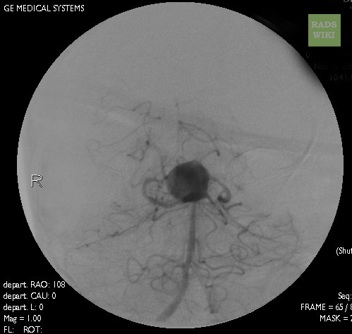
Cranial Angiography: A large basilar artery aneurysm
-
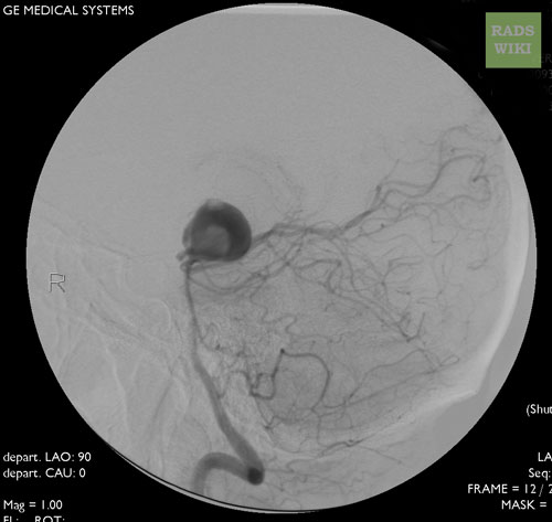
Cranial Angiography: A large basilar artery aneurysm


Complications of Interventions
Images shown below are courtesy of RadsWiki and copylefted
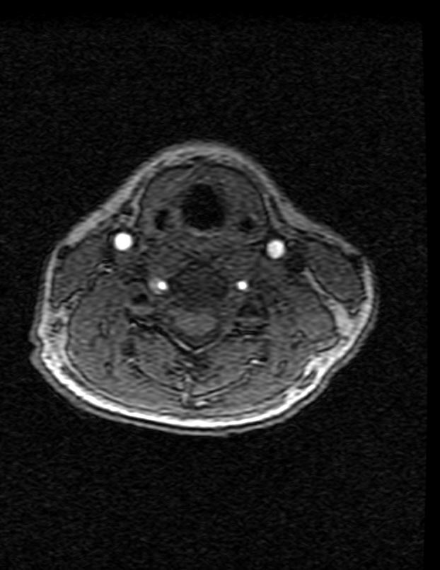
-
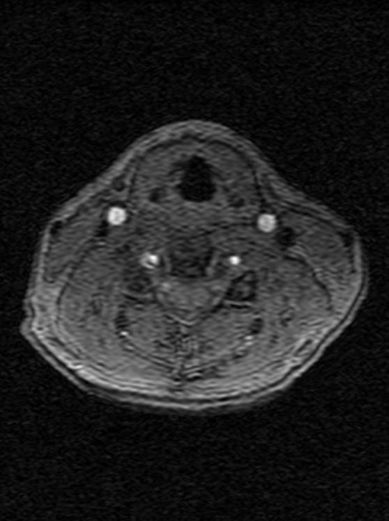
CT: Vertebral artery dissection
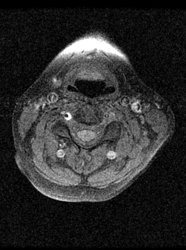
-
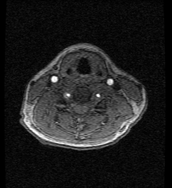
CT: Vertebral artery dissection


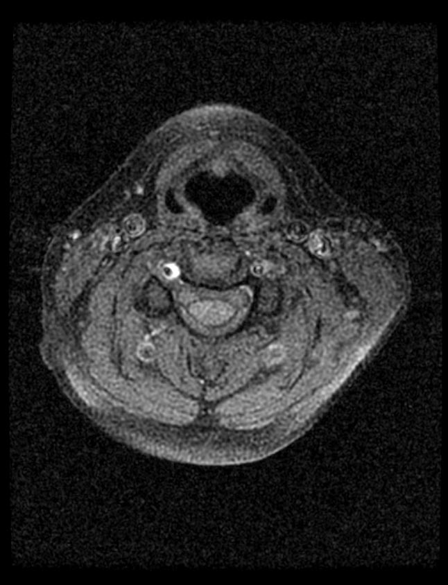
-
CT: Vertebral artery dissection
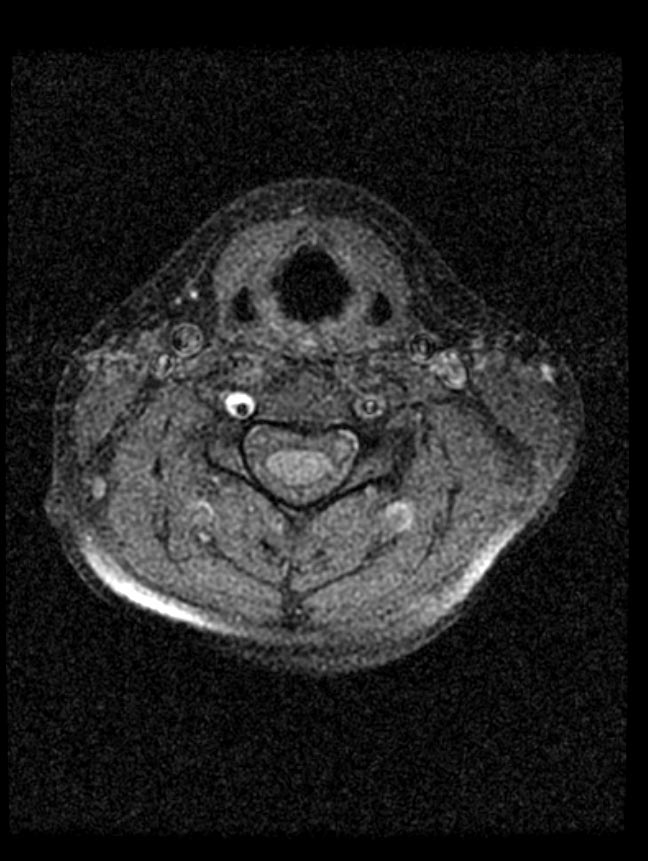
-
CT: Vertebral artery dissection


-
CT: Vertebral artery dissection
-
CT: Vertebral artery dissection
-
CT: Vertebral artery dissection


