Atrial flutter electrocardiogram: Difference between revisions
No edit summary |
|||
| Line 25: | Line 25: | ||
# QRS either normal or aberrant depending on preexisting IVCD or aberrant ventricular conduction. | # QRS either normal or aberrant depending on preexisting IVCD or aberrant ventricular conduction. | ||
===[[EKG Examples of Atrial flutter]]=== | |||
---- | ---- | ||
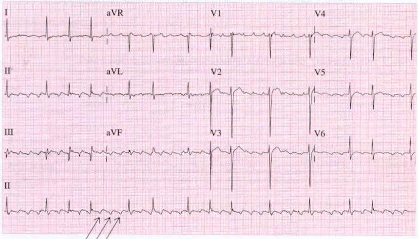
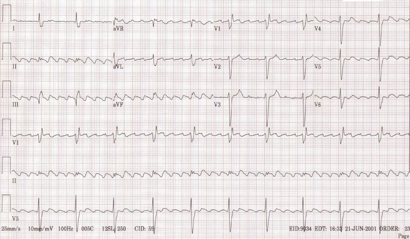
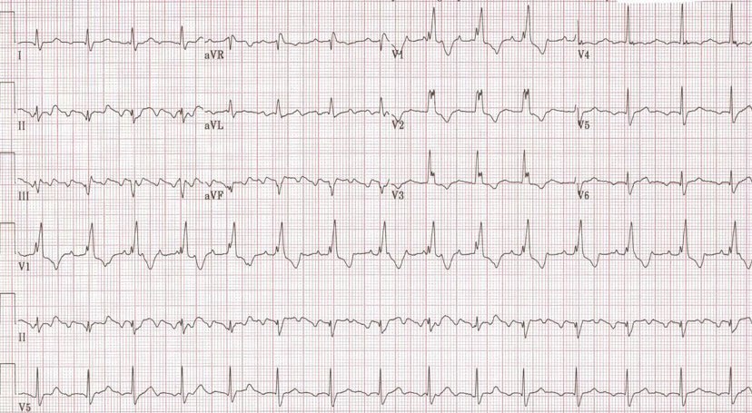
This is a cardiogram from a middle aged man with palpitations. The patient had a dual chamber pacemaker (Vista) set at a lower rate of 60/min, and upper rate of 100/min. The patient had developed atrial flutter, and the pacemaker was following this with a 3:1 block. The flutter is seen in the second panel where the pacemaker was set to a VVI mode and a rate of 30/min. Note that in the first tracing the pacer spikes are not well seen as the pacemaker is pacing in a bipolar mode. | This is a cardiogram from a middle aged man with palpitations. The patient had a dual chamber pacemaker (Vista) set at a lower rate of 60/min, and upper rate of 100/min. The patient had developed atrial flutter, and the pacemaker was following this with a 3:1 block. The flutter is seen in the second panel where the pacemaker was set to a VVI mode and a rate of 30/min. Note that in the first tracing the pacer spikes are not well seen as the pacemaker is pacing in a bipolar mode. | ||
Revision as of 14:17, 15 October 2012
|
Atrial flutter Microchapters |
|
Diagnosis |
|---|
|
Treatment |
|
Case Studies |
|
Atrial flutter electrocardiogram On the Web |
|
American Roentgen Ray Society Images of Atrial flutter electrocardiogram |
|
Risk calculators and risk factors for Atrial flutter electrocardiogram |
Please help WikiDoc by adding content here. It's easy! Click here to learn about editing.
Editor-In-Chief: C. Michael Gibson, M.S., M.D. [1]; Associate Editor-In-Chief: Cafer Zorkun, M.D., Ph.D. [2]
Overview
Electrocardiogram
Electrocardiographic findings are as follows:
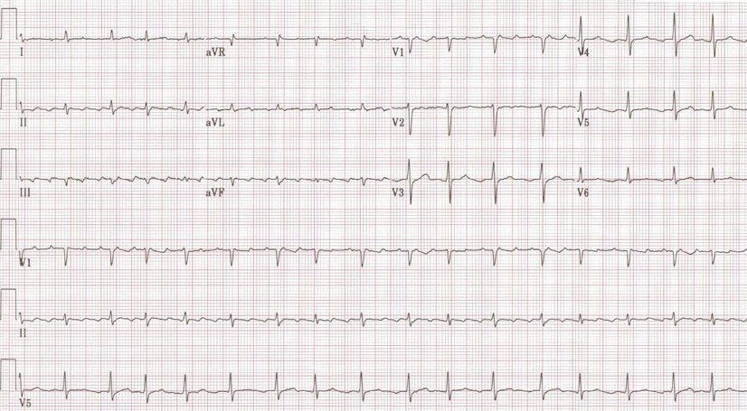
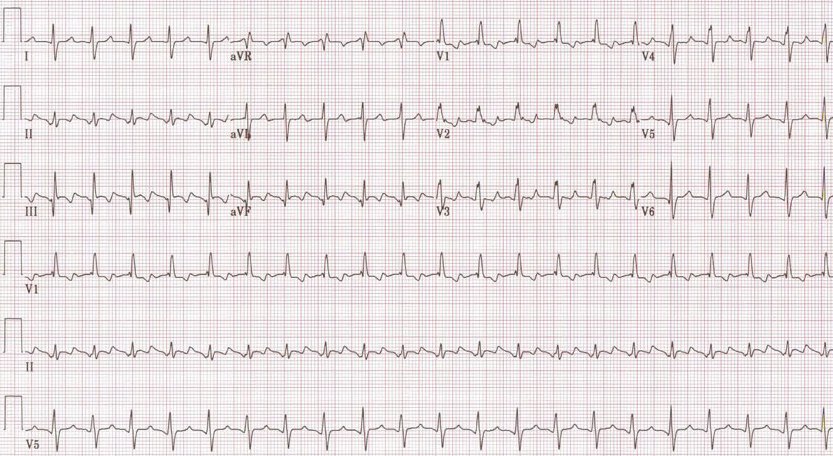
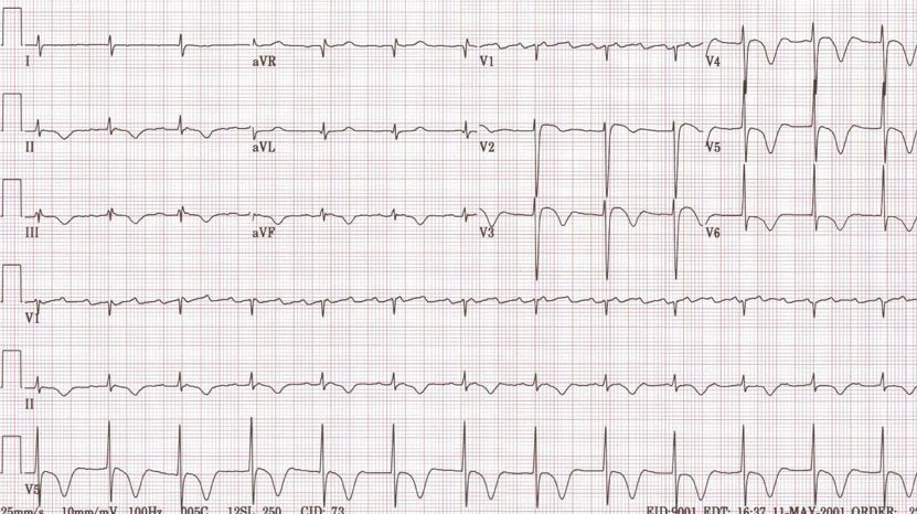
- There are rapid regular undulations (F waves) that cause a sawtooth appearance.
- Best seen in 2,3,F and V1.
- Usually inverted in the inferior leads.
- No isoelectric baselines between the F waves.
- Atrial rate is 250 to 350 Beats Per Minute (BPM).
- Can be faster in infants and children.
- Massive dilation of the atria can lead to a rate < 200 BPM.
- Quinidine can reduce the atrial rate.
- There is a variable ventricular rate depending on the AV conduction.
- The Most common response is 2:1
- 3:1 is uncommon
- 4:1 suggests the existence of an AV conduction defect
- May be associated with complete AV block in which case the RR intervals are regular and the F waves have no constant relationship to the QRS. The ventricular response is usually slow.
- 1:1 conduction may be precipitated by excitement, exercise, induction of anesthesia or any increase in sympathetic tone. It may occur in WPW where the impulses are conducted antegrade through the bypass tract. All these are an emergency.
- During treatment with quinidine the atrial rate may slow sufficiently to permit 1:1 conduction.
- Vagal maneuvers increase the degree of AV block.
- QRS either normal or aberrant depending on preexisting IVCD or aberrant ventricular conduction.
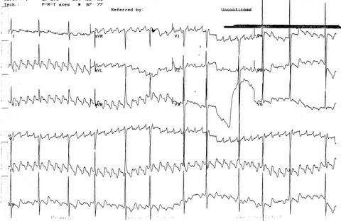
EKG Examples of Atrial flutter
This is a cardiogram from a middle aged man with palpitations. The patient had a dual chamber pacemaker (Vista) set at a lower rate of 60/min, and upper rate of 100/min. The patient had developed atrial flutter, and the pacemaker was following this with a 3:1 block. The flutter is seen in the second panel where the pacemaker was set to a VVI mode and a rate of 30/min. Note that in the first tracing the pacer spikes are not well seen as the pacemaker is pacing in a bipolar mode.
Examples
-
Atrial flutter
-
Atrial flutter with variable conduction
-
A very rare condition 1:1 Atrial flutter
-
2:1 Atrial flutter
-
3:1 Atrial flutter
-
4:1 Atrial flutter
-
Atrial flutter with RBBB
-
Typical atrial flutter pattern


