Anthrax pathophysiology: Difference between revisions
Joao Silva (talk | contribs) No edit summary |
Joao Silva (talk | contribs) No edit summary |
||
| Line 3: | Line 3: | ||
{{CMG}} | {{CMG}} | ||
==Overview== | ==Overview== | ||
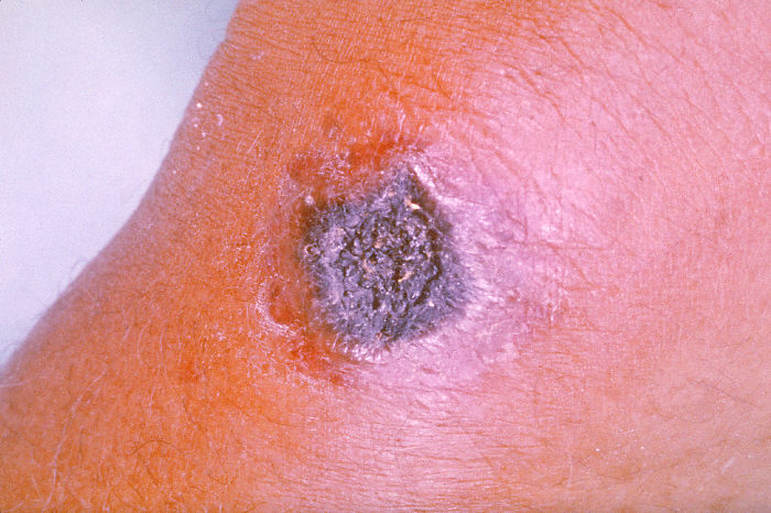
There are 3 routes by which a person can become infected: Inhalation, ingestion and skin exposure. Once Antrhax spores are inhaled they are transported through the air passages into the tiny air sacs ([[alveoli]]) in the lungs. The spores are then picked up by scavenger cells ([[macrophages]]) in the lungs and are transported through small vessels ([[lymphatics]]) to the glands ([[lymph nodes]]) in the central chest cavity ([[mediastinum]]). Damage to the lungs causes [[chest pain]] and [[difficulty breathing]]. Once in the lymph glands, the spores germinate into active bacillus, that multiplies, and eventually bursts the macrophage cell, releasing many more bacilli into the bloodstream which are transferred to the entire body. Once in the blood stream these bacilli release a tripartite toxin (composed of lethal factor, edema factor and protective antigen) which is known to be the primary agents of tissue destruction, bleeding, and death. If antibiotics are given too late, even if the antibiotics eradicate the bacteria, some people still will die because the toxins produced by the bacilli still remain in their system at lethal dose levels. Eating anthrax infected meat is another mode of infection and is characterized by [[vomiting of blood]], severe [[diarrhea]], acute inflammation of the intestinal tract, and [[loss of appetite]]. Gastrointestinal infections can be treated but usually result in fatality rates of 25% to 60%, depending upon how soon treatment commences. Cutaneous (on the skin) anthrax infection causes a boil-like skin lesion that eventually forms an ulcer with a black center (i.e., [[eschar]]). The black eschar often shows up as a large, painless necrotic ulcers (beginning as an irritating and itchy skin lesion or blister that is dark and usually concentrated as a black dot, somewhat resembling bread mold) at the site of infection. Cutaneous infection is the least fatal form of anthrax infection if treated. But without treatment, approximately 20% of all cutaneous skin infection cases may progress to [[toxemia]] and death. <ref>{{cite web | title = Anthrax Q & A: Signs and Symptoms | work = Emergency Preparedness and Response | publisher = Centers for Disease Control and Prevention | date = 2003 | url = http://www.bt.cdc.gov/agent/anthrax/faq/signs.asp | accessdate = 2007-04-19 }}</ref> Treated cutaneous anthrax is rarely fatal.<ref name="bravata"/> | |||
==Transmission== | ==Transmission== | ||
The route by which anthrax is transmitted allows for its classification, it includes: | |||
* Cutaneous anthrax - commonly held to require a prior skin lesion as a prerequisite for infection | |||
* Gastrointestinal anthrax - contracted following ingestion of contaminated food, primarily meat from an animal that died of the disease, or conceivably from ingestion of contami- nated water | |||
* Inhalational anthrax - from breathing in airborne anthrax spores | |||
* Injection anthrax - from injection of a drug containing or contaminated with Bacillus anthracis | |||
==Genetics== | ==Genetics== | ||
Revision as of 15:39, 18 July 2014
|
Anthrax Microchapters |
|
Diagnosis |
|---|
|
Treatment |
|
Case Studies |
|
Anthrax pathophysiology On the Web |
|
American Roentgen Ray Society Images of Anthrax pathophysiology |
|
Risk calculators and risk factors for Anthrax pathophysiology |
Editor-In-Chief: C. Michael Gibson, M.S., M.D. [1]
Overview
There are 3 routes by which a person can become infected: Inhalation, ingestion and skin exposure. Once Antrhax spores are inhaled they are transported through the air passages into the tiny air sacs (alveoli) in the lungs. The spores are then picked up by scavenger cells (macrophages) in the lungs and are transported through small vessels (lymphatics) to the glands (lymph nodes) in the central chest cavity (mediastinum). Damage to the lungs causes chest pain and difficulty breathing. Once in the lymph glands, the spores germinate into active bacillus, that multiplies, and eventually bursts the macrophage cell, releasing many more bacilli into the bloodstream which are transferred to the entire body. Once in the blood stream these bacilli release a tripartite toxin (composed of lethal factor, edema factor and protective antigen) which is known to be the primary agents of tissue destruction, bleeding, and death. If antibiotics are given too late, even if the antibiotics eradicate the bacteria, some people still will die because the toxins produced by the bacilli still remain in their system at lethal dose levels. Eating anthrax infected meat is another mode of infection and is characterized by vomiting of blood, severe diarrhea, acute inflammation of the intestinal tract, and loss of appetite. Gastrointestinal infections can be treated but usually result in fatality rates of 25% to 60%, depending upon how soon treatment commences. Cutaneous (on the skin) anthrax infection causes a boil-like skin lesion that eventually forms an ulcer with a black center (i.e., eschar). The black eschar often shows up as a large, painless necrotic ulcers (beginning as an irritating and itchy skin lesion or blister that is dark and usually concentrated as a black dot, somewhat resembling bread mold) at the site of infection. Cutaneous infection is the least fatal form of anthrax infection if treated. But without treatment, approximately 20% of all cutaneous skin infection cases may progress to toxemia and death. [1] Treated cutaneous anthrax is rarely fatal.[2]
Transmission
The route by which anthrax is transmitted allows for its classification, it includes:
- Cutaneous anthrax - commonly held to require a prior skin lesion as a prerequisite for infection
- Gastrointestinal anthrax - contracted following ingestion of contaminated food, primarily meat from an animal that died of the disease, or conceivably from ingestion of contami- nated water
- Inhalational anthrax - from breathing in airborne anthrax spores
- Injection anthrax - from injection of a drug containing or contaminated with Bacillus anthracis
Genetics
Pathogenesis
B. anthracis, the causative agent of anthrax, is a spore-forming bacterium. The spores of B. anthracis, which can remain dormant in the environment for decades, are the infectious form, but vegetative B. anthracis rarely causes disease.[3] The bacterium causes disease through 2 mechanisms: toxemia and bacterial infection.[4] Spores introduced through the skin lead to cutaneous or injection anthrax; those introduced through the gastrointestinal tract lead to gastrointestinal anthrax; and those introduced through the lungs lead to inhalation anthrax. After entering a human or animal, B. anthracis spores are believed to germinate locally or be transported by phagocytic cells to the lymphatics and regional lymph nodes, where they germinate; or both.[5] B. anthracis begins producing toxins within hours of germination.[6] Protective antigen (PA) and edema factor (EF) combine to form edema toxin (ET) and PA and lethal factor (LF) combine to form lethal toxin (LT). After binding to surface receptors, the PA portion of the complexes facilitates translocation of the toxins to the cytosol, in which EF and LF exert their toxic effects.[7]
Gross Pathology
Inhalation Anthrax
Gross pathologic lesions observed in non-human primates used in aerosol challenge models of inhalation anthrax include edema, congestion, hemorrhage and necrosis in the lungs and mediastinum. Splenitis and necrotizing or hemorrhagic lymphadenitis involving the mediastinal, tracheobronchial, and other lymph nodes are common.[8] Primary pulmonary lesions, including those of pneumonia, are occasionally observed. Meningeal involvement ranging from edema, congestion, hemorrhage, and necrosis to suppurative or hemorrhagic meningitis, usually secondary to hematogenous spread from other types of anthrax, occurs in ≤77% of animals studied.[9] Autopsy findings for persons who died from inhalation anthrax in Sverdlovsk and in the United States[10] are consistent with findings from the non-human primates studies. Persons who died had extensive amounts of serosanguinous fluid in pleural cavities and edema and hemorrhage of the mediastinum and surrounding soft tissues, and 48% had cerebral edema, 21% had ascites, 17% had pericardial effusions, and 14% had petechial rash. Mediastinal lymph nodes and spleen also showed hemorrhage and necrosis.[8][11]
Microscopic Pathology
Mode of infection
Anthrax can enter the human body through the intestines (ingestion), lungs (inhalation), or skin (cutaneous) and causes distinct clinical symptoms based on its site of entry. An infected human will generally be quarantined. However, anthrax does not usually spread from an infected human to a noninfected human. But if the disease is fatal the person’s body and its mass of anthrax bacilli becomes a potential source of infection to others and special precautions should be used to prevent more contamination. Unfortunately inhalation anthrax, if left untreated until obvious symptoms occur, will usually result in death if treatment is started too late.
Anthrax is usually contracted by handling infected animals or their wool, germ warfare/terrorism or laboratory accidents.
1) Pulmonary (pneumonic, respiratory, or inhalation) anthrax Respiratory infection initially presents with cold or flu-like symptoms for several days, followed by severe (and often fatal) respiratory collapse. If not treated promptly soon after exposure, before symptoms appear, inhalational anthrax is highly fatal, with near 100% mortality.[2] A lethal dose of anthrax is reported to result from inhalation of about 10,000–20,000 spores. [12] Like all diseases there is probably a wide variation to susceptibility with evidence that some people may die from much lower exposures; there is little documented evidence to verify the exact or average number of spores need for infection. Inhalation anthrax is also known as Woolsorters' disease or as Ragpickers' disease since these people often caught it. Other practices associated with exposure include the slicing up of animal horns for the manufacture of buttons, the handling of hair bristles used for the manufacturing of brushes, and the handling of animal skins. Whether these animal skins came from animals that died of the disease or from animals that had simply laid on ground that had spores on it is unknown. Anthrax is a very hard disease to eliminate since Anthrax spores are devilishly hard to kill and have been known to have reinfected animals over 70 years after burial sites of anthrax infected animals were disturbed. [13]
2) Gastrointestinal (gastroenteric) anthrax Gastrointestinal infection is most often caused by eating anthrax infected meat and is characterized by serious gastrointestinal difficulty, vomiting of blood, severe diarrhea, acute inflammation of the intestinal tract, and loss of appetite. Gastrointestinal infections can be treated but usually result in fatality rates of 25% to 60%, depending upon how soon treatment commences. [14]
3) Cutaneous (skin) anthrax

Cutaneous (on the skin) anthrax infection shows up as a boil-like skin lesion that eventually forms an ulcer with a black center (i.e., eschar). The black eschar often shows up as a large, painless necrotic ulcers (beginning as an irritating and itchy skin lesion or blister that is dark and usually concentrated as a black dot, somewhat resembling bread mold) at the site of infection. Cutaneous infections generally form within the site of spore penetration within 2 to 5 days after exposure. Unlike bruises or most other lesions, cutaneous anthrax infections normally do not cause pain. Cutaneous infection is the least fatal form of anthrax infection if treated. But without treatment, approximately 20% of all cutaneous skin infection cases may progress to toxemia and death. [15] Treated cutaneous anthrax is rarely fatal.[2]
References
- ↑ "Anthrax Q & A: Signs and Symptoms". Emergency Preparedness and Response. Centers for Disease Control and Prevention. 2003. Retrieved 2007-04-19.
- ↑ 2.0 2.1 2.2 Bravata DM, Holty JE, Liu H, McDonald KM, Olshen RA, Owens DK (2006), Systematic review: a century of inhalation anthrax cases from 1900 to 2005, Annals of Internal Medicine; 144(4): 270–80.
- ↑ Shadomy, Sean V.; Smith, Theresa L. (2008). "Anthrax". Journal of the American Veterinary Medical Association. 233 (1): 63–72. doi:10.2460/javma.233.1.63. ISSN 0003-1488.
- ↑ Liu, Shihui; Moayeri, Mahtab; Leppla, Stephen H. (2014). "Anthrax lethal and edema toxins in anthrax pathogenesis". Trends in Microbiology. 22 (6): 317–325. doi:10.1016/j.tim.2014.02.012. ISSN 0966-842X.
- ↑ Ross, Joan M. (1957). "The pathogenesis of anthrax following the administration of spores by the respiratory route". The Journal of Pathology and Bacteriology. 73 (2): 485–494. doi:10.1002/path.1700730219. ISSN 0368-3494.
- ↑ Hanna, Philip C.; Ireland, John A.W. (1999). "Understanding Bacillus anthracis pathogenesis". Trends in Microbiology. 7 (5): 180–182. doi:10.1016/S0966-842X(99)01507-3. ISSN 0966-842X.
- ↑ Moayeri, M (2004). "The roles of anthrax toxin in pathogenesis". Current Opinion in Microbiology. 7 (1): 19–24. doi:10.1016/j.mib.2003.12.001. ISSN 1369-5274.
- ↑ 8.0 8.1 Guarner, Jeannette; Jernigan, John A.; Shieh, Wun-Ju; Tatti, Kathleen; Flannagan, Lisa M.; Stephens, David S.; Popovic, Tanja; Ashford, David A.; Perkins, Bradley A.; Zaki, Sherif R. (2003). "Pathology and Pathogenesis of Bioterrorism-Related Inhalational Anthrax". The American Journal of Pathology. 163 (2): 701–709. doi:10.1016/S0002-9440(10)63697-8. ISSN 0002-9440.
- ↑ Twenhafel, N. A. (2010). "Pathology of Inhalational Anthrax Animal Models". Veterinary Pathology. 47 (5): 819–830. doi:10.1177/0300985810378112. ISSN 0300-9858.
- ↑ A. A. Abramova & L. M. Grinberg (1993). "[Pathology of anthrax sepsis according to materials of the infectious outbreak in 1979 in Sverdlovsk (macroscopic changes)]". Arkhiv patologii. 55 (1): 12–17. PMID 7980032. Unknown parameter
|month=ignored (help) - ↑ A. A. Abramova & L. M. Grinberg (1993). "[Pathology of anthrax sepsis according to materials of the infectious outbreak in 1979 in Sverdlovsk (macroscopic changes)]". Arkhiv patologii. 55 (1): 12–17. PMID 7980032. Unknown parameter
|month=ignored (help) - ↑ "www.medicinenet.com". Retrieved 2012-08-31.
- ↑ "Anthrax" by Jeanne Guillemin, University of California Press, 2001,ISBN 0-520-22917-7, pg. 3
- ↑ "Anthrax Q & A: Signs and Symptoms". Emergency Preparedness and Response. Centers for Disease Control and Prevention. 2003. Retrieved 2007-04-19.
- ↑ "Anthrax Q & A: Signs and Symptoms". Emergency Preparedness and Response. Centers for Disease Control and Prevention. 2003. Retrieved 2007-04-19.