Abdominal aortic aneurysm CT: Difference between revisions
(New page: {{Template:Abdominal aortic aneurysm}} {{CMG}} '''Associate Editor-In-Chief:''' {{CZ}} === Contrast CT === * Provides detailed anatomic information and is valuable in planning AAA repa...) |
m (Bot: Adding CME Category::Cardiology) |
||
| (15 intermediate revisions by 6 users not shown) | |||
| Line 1: | Line 1: | ||
__NOTOC__ | |||
{{Template:Abdominal aortic aneurysm}} | {{Template:Abdominal aortic aneurysm}} | ||
{{CMG}}; '''Associate Editor-In-Chief:''' {{CZ}} | |||
==Overview== | |||
Although [[CT]] scanning provides detailed anatomic information and is valuable in planning abdominal aortic aneurysm repair, it is not used as a screening tool because of the potential [[nephrotoxicity]] associated with the dye load, the cost, and the exposure to radiation. | |||
==Limitations of CT Scanning== | |||
The limitations include: | |||
*Potential [[nephrotoxicity]] associated with the dye load | |||
*Cost | |||
*Exposure to radiation | |||
*Suboptimal visualization of the origins of the aortic branch vessels | |||
*Occasionally, inaccurate localization of the aneurysmal neck<ref name="Ernst-1993">{{Cite journal | last1 = Ernst | first1 = CB. | title = Abdominal aortic aneurysm. | journal = N Engl J Med | volume = 328 | issue = 16 | pages = 1167-72 | month = Apr | year = 1993 | doi = 10.1056/NEJM199304223281607 | PMID = 8455684 }}</ref> | |||
== | ==CT Examples== | ||
[http://www.peir.net Image courtesy of Professor Peter Anderson DVM PhD and published with permission © PEIR, University of Alabama at Birmingham, Department of Pathology]: | |||
<div align="left"> | |||
<gallery heights="175" widths="175"> | |||
Image:Abdominal Aortic Aneurysm 0001.jpg|Abdominal Aortic Aneurysm | |||
Image:Abdominal Aortic Aneurysm 0002.jpg|Abdominal Aortic Aneurysm | |||
</gallery> | |||
</div> | |||
<div align="left"> | |||
<gallery heights="175" widths="175"> | |||
Image:Abdominal Aortic Aneurysm 0003.jpg|Abdominal Aortic Aneurysm | |||
Image:Abdominal Aortic Aneurysm 0004.jpg|Abdominal Aortic Aneurysm | |||
</gallery> | |||
</div> | |||
<div align="left"> | |||
<gallery heights="175" widths="175"> | |||
Image:Abdominal Aortic Aneurysm 0005.jpg|Abdominal Aortic Aneurysm | |||
Image:Abdominal Aortic Aneurysm 0006.jpg|Abdominal Aortic Aneurysm | |||
</gallery> | |||
</div> | |||
<div align="left"> | |||
<gallery heights="175" widths="175"> | |||
Image:Abdominal Aortic Aneurysm 0007.jpg|Abdominal Aortic Aneurysm | |||
Image:Abdominal Aortic Aneurysm 0008.jpg|Abdominal Aortic Aneurysm | |||
</gallery> | |||
</div> | |||
Copyleft image obtained courtesy of Radswiki: | |||
<div align="left"> | |||
<gallery heights="175" widths="175"> | |||
Image:Abdominal aortic aneurysm 001.jpg|CT: a large abdominal aortic aneurysm | |||
Image:Abdominal aortic aneurysm 002.jpg|CT: a large abdominal aortic aneurysm | |||
Image:Abdominal aortic aneurysm 003.jpg|CT: a large abdominal aortic aneurysm | |||
</gallery> | |||
</div> | |||
<div align="left"> | |||
<gallery heights="175" widths="175"> | |||
Image:Abdominal aortic aneurysm 101.jpg|CT: a large abdominal aortic aneurysm | |||
Image:Abdominal aortic aneurysm 102.jpg|CT: a large abdominal aortic aneurysm | |||
</gallery> | |||
</div> | |||
<div align="left"> | |||
<gallery heights="175" widths="175"> | |||
Image:Abdominal aortic aneurysm 103.jpg|CT: a large abdominal aortic aneurysm | |||
Image:Abdominal aortic aneurysm 104.jpg|CT: a large abdominal aortic aneurysm | |||
</gallery> | |||
</div> | |||
<div align="left"> | |||
<gallery heights="175" widths="175"> | |||
Image:Ruptured abdominal aortic aneurysm 001.jpg|Ruptured abdominal aortic aneurysm | |||
Image:Ruptured abdominal aortic aneurysm 002.jpg|Ruptured abdominal aortic aneurysm | |||
</gallery> | |||
</div> | |||
<div align="left"> | |||
<gallery heights="175" widths="175"> | |||
Image:Ruptured abdominal aortic aneurysm 003.jpg|Ruptured abdominal aortic aneurysm | |||
Image:Ruptured aaa.jpg|This patient presented with acute abdominal pain and hypotension. His non-contrast CT shows a large AAA and extensive periaortic haematoma. A thick (but subtle) hyperdense crescent is present within the aortic wall posteriorly and laterally which represents acute intramural hematoma, a sign of acute or impending rupture. (Image courtesy of Dr Donna D'Souza) | |||
</gallery> | |||
</div> | |||
==References== | ==References== | ||
{{Reflist|2}} | {{Reflist|2}} | ||
{{WH}} | |||
{{WS}} | |||
[[CME Category::Cardiology]] | |||
[[Category: | [[Category:Disease]] | ||
[[Category:Cardiac surgery]] | [[Category:Cardiac surgery]] | ||
[[Category:Surgery]] | [[Category:Surgery]] | ||
[[Category:Cardiology]] | [[Category:Cardiology]] | ||
[[Category:Up-To-Date cardiology]] | |||
[[Category:Up-To-Date]] | |||
[[Category:Best pages]] | |||
Latest revision as of 18:23, 14 March 2016
|
Abdominal Aortic Aneurysm Microchapters |
|
Differentiating Abdominal Aortic Aneurysm from other Diseases |
|---|
|
Diagnosis |
|
Treatment |
|
Case Studies |
|
Abdominal aortic aneurysm CT On the Web |
|
Directions to Hospitals Treating Abdominal aortic aneurysm CT |
|
Risk calculators and risk factors for Abdominal aortic aneurysm CT |
Editor-In-Chief: C. Michael Gibson, M.S., M.D. [1]; Associate Editor-In-Chief: Cafer Zorkun, M.D., Ph.D. [2]
Overview
Although CT scanning provides detailed anatomic information and is valuable in planning abdominal aortic aneurysm repair, it is not used as a screening tool because of the potential nephrotoxicity associated with the dye load, the cost, and the exposure to radiation.
Limitations of CT Scanning
The limitations include:
- Potential nephrotoxicity associated with the dye load
- Cost
- Exposure to radiation
- Suboptimal visualization of the origins of the aortic branch vessels
- Occasionally, inaccurate localization of the aneurysmal neck[1]
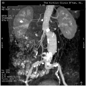
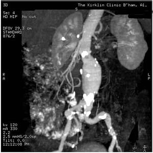
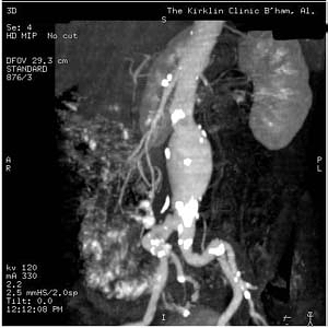
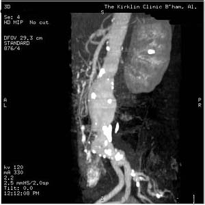
CT Examples
-
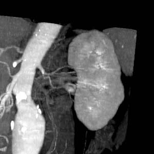
Abdominal Aortic Aneurysm
-
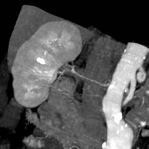
Abdominal Aortic Aneurysm


-
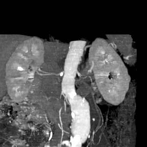
Abdominal Aortic Aneurysm
-
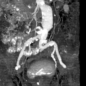
Abdominal Aortic Aneurysm


-
Abdominal Aortic Aneurysm
-
Abdominal Aortic Aneurysm


-
Abdominal Aortic Aneurysm
-
Abdominal Aortic Aneurysm


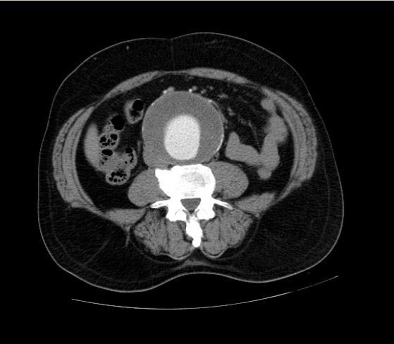
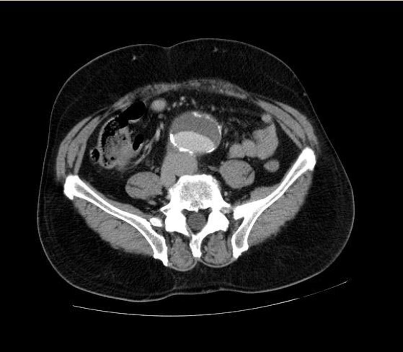
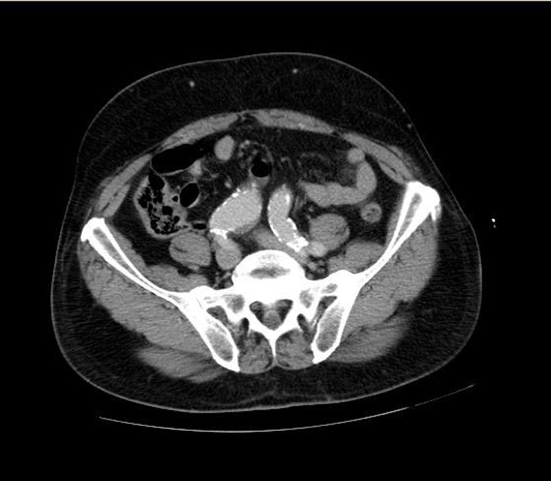
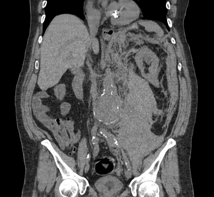
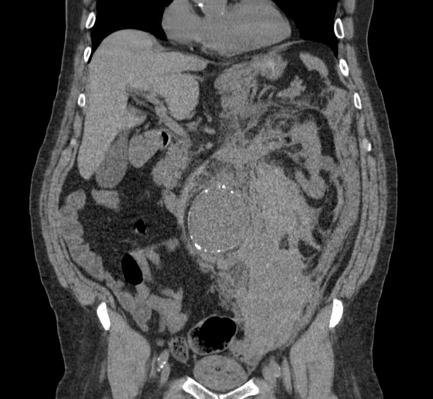
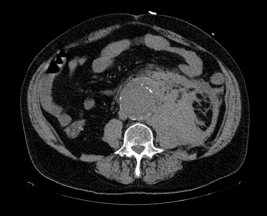
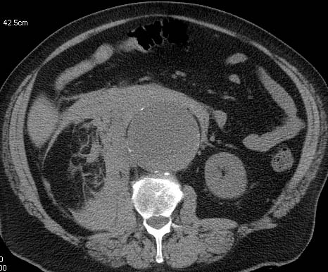
Copyleft image obtained courtesy of Radswiki:
-
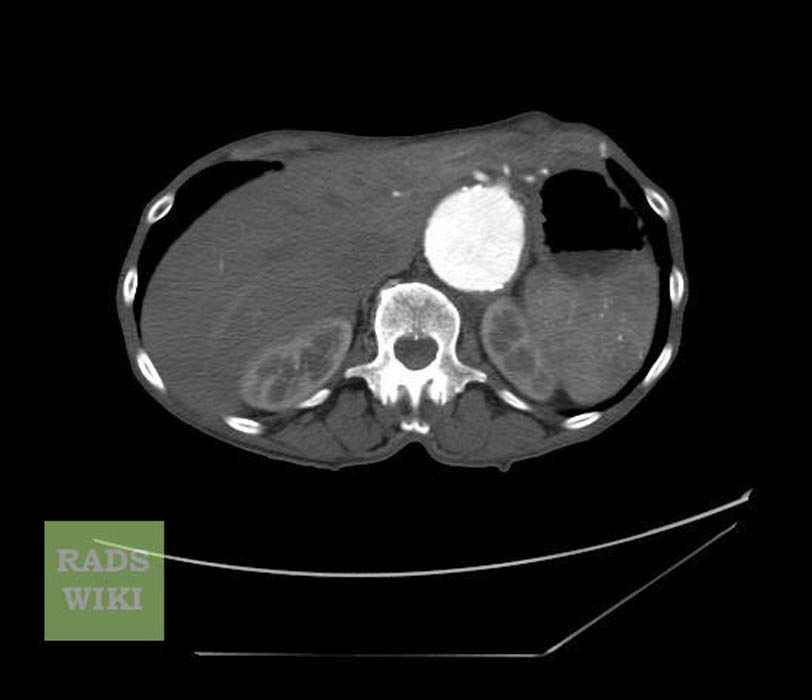
CT: a large abdominal aortic aneurysm
-
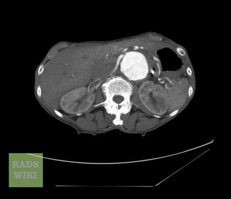
CT: a large abdominal aortic aneurysm
-
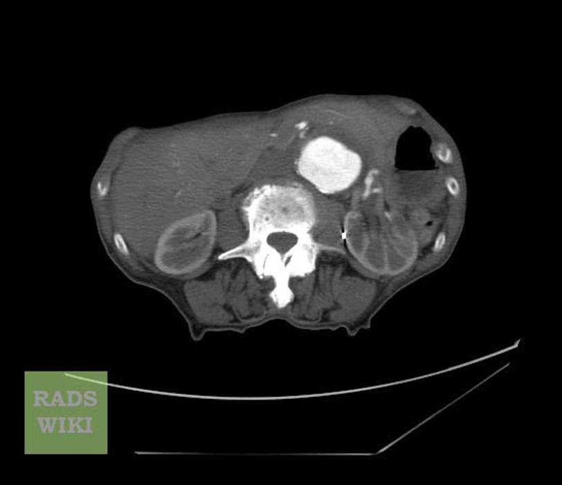
CT: a large abdominal aortic aneurysm


-
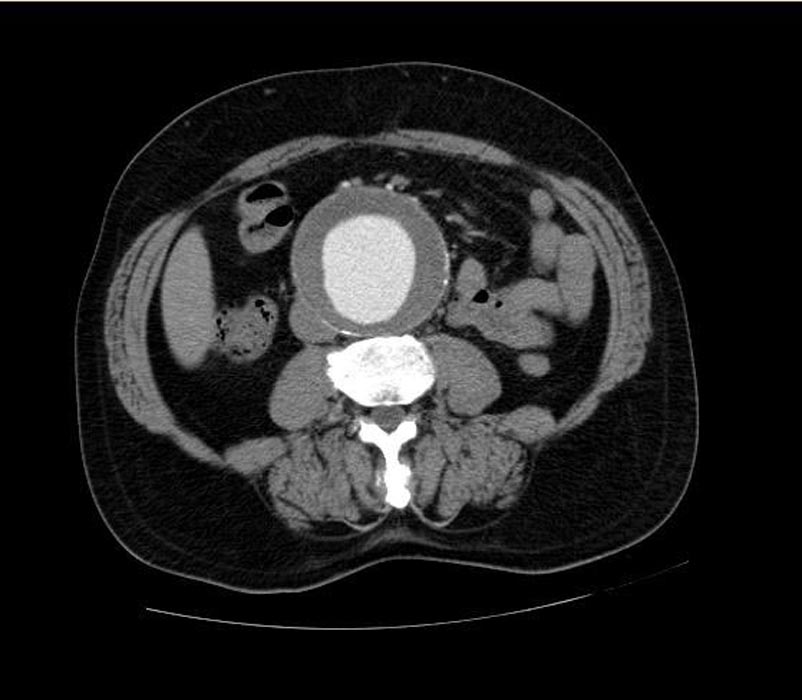
CT: a large abdominal aortic aneurysm
-
CT: a large abdominal aortic aneurysm


-
CT: a large abdominal aortic aneurysm
-
CT: a large abdominal aortic aneurysm


-
Ruptured abdominal aortic aneurysm
-
Ruptured abdominal aortic aneurysm


-
Ruptured abdominal aortic aneurysm
-
This patient presented with acute abdominal pain and hypotension. His non-contrast CT shows a large AAA and extensive periaortic haematoma. A thick (but subtle) hyperdense crescent is present within the aortic wall posteriorly and laterally which represents acute intramural hematoma, a sign of acute or impending rupture. (Image courtesy of Dr Donna D'Souza)

