Premature Ventricular Contraction EKG Examples
Editor-In-Chief: C. Michael Gibson, M.S., M.D. [1]
For the main page on premature ventricular contraction, click here.
Premature Ventricular Contraction EKG Examples
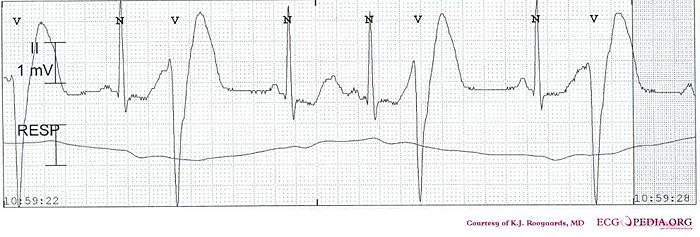
Shown below is an EKG showing premature ventricular contractions.

Copyleft image obtained courtesy of ECGpedia, http://en.ecgpedia.org/wiki/File:De-KJcasu17-1.jpg
Shown below is an EKG showing a Premature Ventricular Contractions.

Copyleft image obtained courtesy of ECGpedia, http://en.ecgpedia.org/wiki/Main_Page
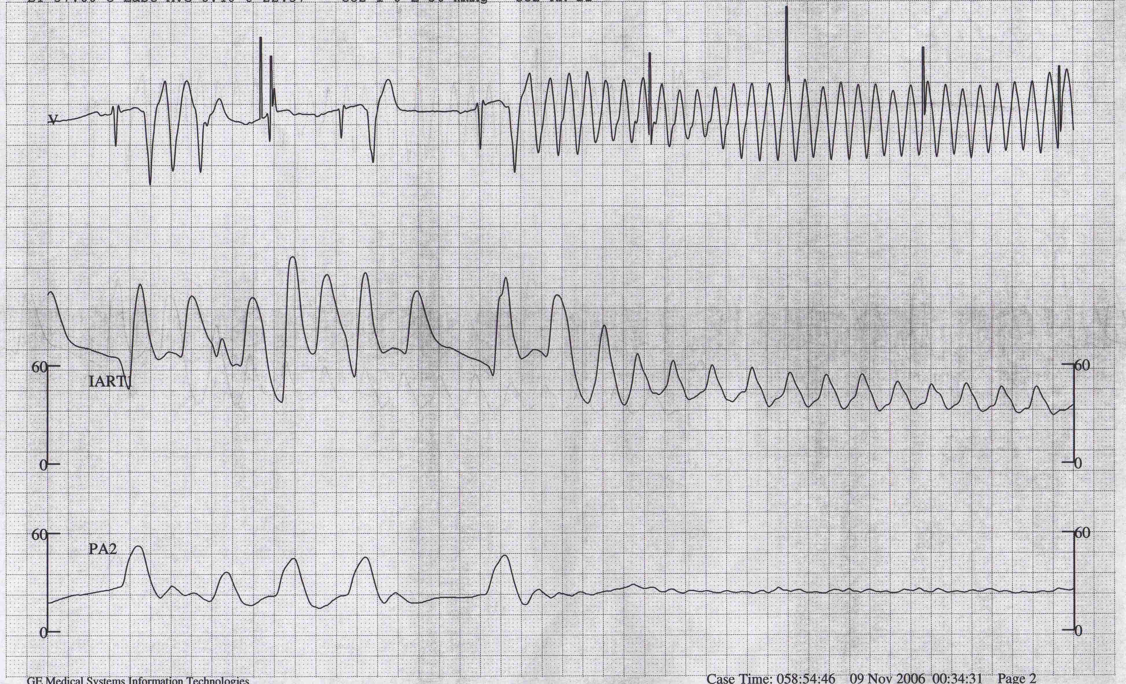
Shown below is an EKG showing a Premature Ventricular Contractions triggering ventricular flutter.

Copyleft image obtained courtesy of ECGpedia, http://en.ecgpedia.org/wiki/Main_Page
Shown below is an EKG showing a sinus rhythm with ventricular bigemini.

Copyleft image obtained courtesy of ECGpedia, http://en.ecgpedia.org/wiki/File:E284.jpg
Shown below is an EKG showing a sinus rhythm with ventricular bigemini. Every sinus beat is followed by a ventricular extrasystole.

Copyleft image obtained courtesy of ECGpedia, http://en.ecgpedia.org/wiki/File:Rhythm_bigemini.png
Shown below is an EKG image of premature ventricular contraction. The arrow indicates a ventricular extrasystole (VES).

Shown below is an EKG showing a trigeminal rhythm where wide complex beats are interpolated between normal QRS complexes. Note that the PR interval is prolonged after the wide complex beat. The wide complex beats are probably ventricular beats. As a rule interpolated wide beats are usually ventricular. In most cases the P wave after the PVC is not conducted and a compensatory pause is created instead of an interpolated beat.

EKG below shows sinus rhythm with interpolated ventricular premature complexes. Wide complex beats interpolated between sinus complexes suggests strongly that they are ventricular in origin.

References