Brugada syndrome electrocardiogram
|
Brugada syndrome Microchapters |
|
Diagnosis |
|---|
|
Treatment |
|
Case Studies |
|
Brugada syndrome electrocardiogram On the Web |
|
American Roentgen Ray Society Images of Brugada syndrome electrocardiogram |
|
Risk calculators and risk factors for Brugada syndrome electrocardiogram |
Editor-In-Chief: C. Michael Gibson, M.S., M.D. [1]
Overview
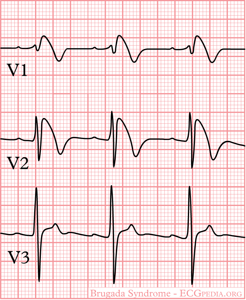
In some cases, the disease can be detected by observing characteristic patterns on an electrocardiogram, which may be present all the time, or might be elicited by the administration of particular drugs (e.g., Class IC antiarrythmic drugs that blocks sodium channels and causing appearance of ECG abnormalities - ajmaline, flecainide) or resurface spontaneously due to as yet unclarified triggers. The pattern seen on the ECG is persistent ST elevations in the electrocardiographic leadsV1-V3 with a right bundle branch block (RBBB) appearance with or without the terminal S waves in the lateral leads that are associated with a typical RBBB. A prolongation of the PR interval (a conduction disturbance in the heart) is also frequently seen.The electrocardiogram can fluctuate over time, depending on the autonomic balance and the administration of antiarrhythmic drugs. Adrenergic stimulation decreases the ST segment elevation, while vagal stimulation worsens it. (There is a case report of a patient who died while shaving, presumed due to the vagal stimulation of the carotid sinus massage!) The administration of class Ia, Ic and III drugs increases the ST segment elevation, and also fever. Exercise decreases ST segment elevation in some patients but increases it in others (after exercise when the body temperature has risen). The changes in heart rate induced by atrial pacing are accompanied by changes in the degree of ST segment elevation. When the heart rate decreases, the ST segment elevation increases and when the heart rate increases the ST segment elevation decreases. However, the contrary can also be observed.
Characteristics
- Characterized by a coved-type ST-segment elevation in the right precordial leads
- The Brugada ECG is often concealed, but can be unmasked or modulated by a number of drugs and pathophysiological states including sodium channel blockers, a febrile state, vagotonic agents, tricyclic antidepressants, as well as cocaine and Propranolol intoxication.
EKG Images
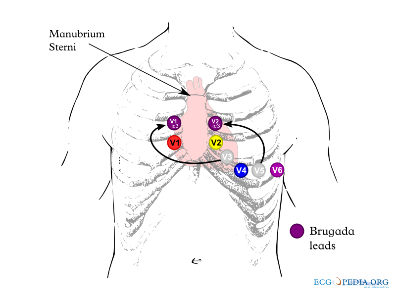
Shown below is an example of lead placements in Brugada syndrome.

Shown below is an example of general EKG characteristics in Brugada syndrome - right bundle branch block in leads V1-2 and ST segment elevation in leads V1-3

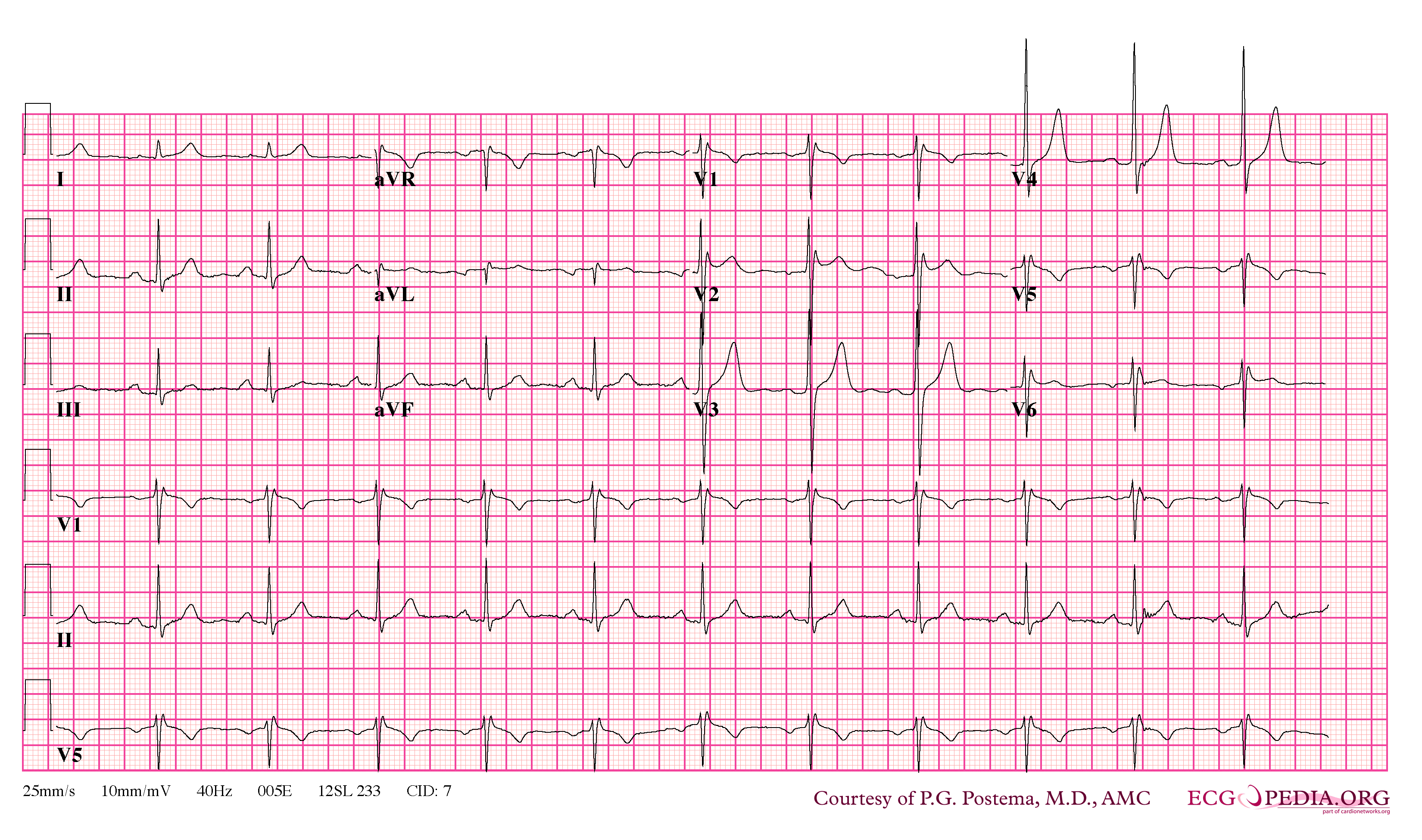
Shown below is an example of Brugada syndrome with ST segment elevation.

Shown below are examples of Brugada syndrome type 1 showing J point elevation, right bundle branch block and T wave inversion.


Shown below are examples of Brugada sydrome type 2 showing J point elevation, saddle shaped ST segment.


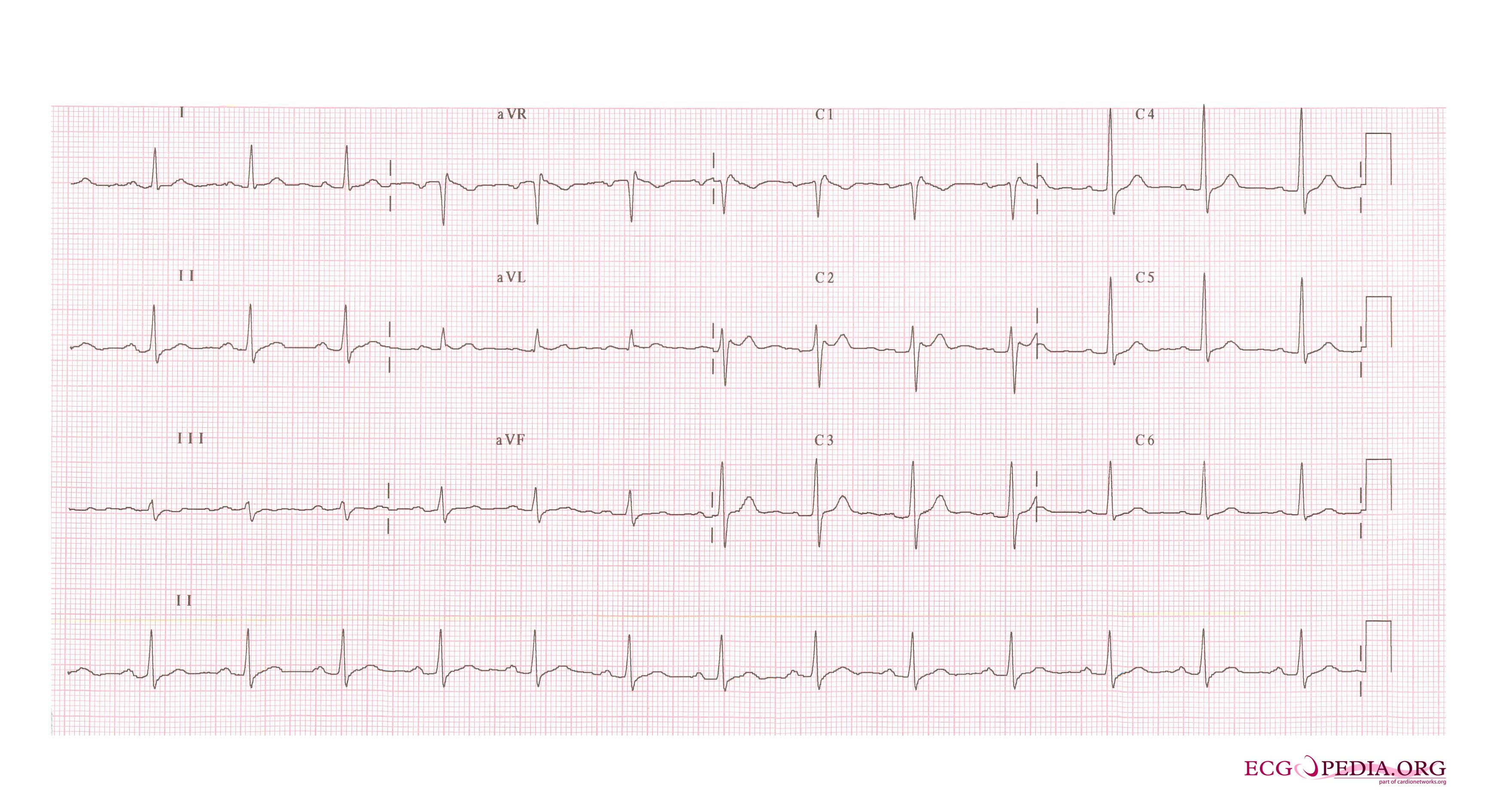
Shown below is an example of Brugada syndrome with right bundle branch block in V1 and St segment elevation in V1-3
