Tuberculosis CT
|
Tuberculosis Microchapters |
|
Diagnosis |
|---|
|
Treatment |
|
Case Studies |
|
Tuberculosis CT On the Web |
|
American Roentgen Ray Society Images of Tuberculosis CT |
Editor-In-Chief: C. Michael Gibson, M.S., M.D. [1]; Associate Editor(s)-in-Chief: Alejandro Lemor, M.D. [2]
Overview
The majority of patients with pulmonary tuberculosis will have abnormal findings in a chest CT, which include micronodules, interlobular septal thickening, cavitation and consolidation.
Computed Tomography
Pulmonary Tuberculosis
- Chest CT abnormalities are seen in the majority of patients with active pulmonary tuberculosis.
- CT finding include:[1]
- Micronodules
- Interlobular septal thickening
- Cavitation
- Consolidation.
- Micronodules are most commonly located in the subpleural region and peribronchovascular interstitium.
Extrapulmonary Tuberculosis
Cardiac Tuberculosis
- Pericardial thickening may be seen in a CT, specially if it is more than 3 mm.[2]
- Lymph node enlargment is also a common CT findings in cardiac tuberculosis.[2]
- Pericardial effusion is seen in less than 20% of patients.[2]
Disseminated Tuberculosis
 |
 |
 |
Tuberculous Meningitis
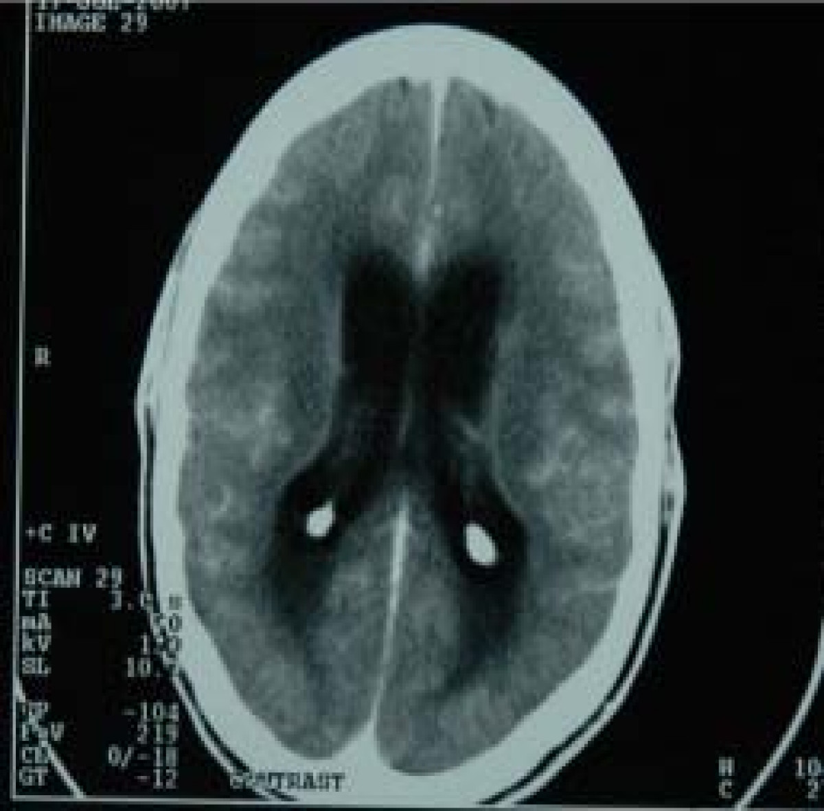
- Head CT findings in tuberculous meningitis include meningeal enhancement consistent with meningeal inflammation and coroidal calcifications.[3]
- Areas of infarction and hemorrhage may also be seen in cases of miliar tuberculosis.
- Patients with late complications may show hydrocephalus.

Abdominal Tuberculosis
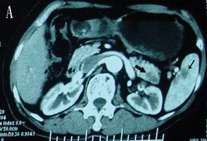
- CT findings in a pancreatic and spleen infection with tuberculosis may mimic a pancreatic cancer.[4]
- Shown below there is CT scan of the pancreas demonstrating a mass in the pancreatic tail and metastasizes in the spleen.
 |
 |
References
- ↑ Jeong Min Ko, Hyun Jin Park & Chi Hong Kim (2014). "Pulmonary Changes of Pleural Tuberculosis: Up-to-Date CT Imaging". Chest. doi:10.1378/chest.14-0196. PMID 25086249. Unknown parameter
|month=ignored (help) - ↑ 2.0 2.1 2.2 Burrill, Joshua; Williams, Christopher J.; Bain, Gillian; Conder, Gabriel; Hine, Andrew L.; Misra, Rakesh R. (2007). "Tuberculosis: A Radiologic Review1". RadioGraphics. 27 (5): 1255–1273. doi:10.1148/rg.275065176. ISSN 0271-5333.
- ↑ Komolafe, Morenikeji A; Sunmonu, Taofiki A; Esan, Olufunmi A (2008). "Tuberculous meningitis presenting with unusual clinical features in Nigerians: Two case reports". Cases Journal. 1 (1): 180. doi:10.1186/1757-1626-1-180. ISSN 1757-1626.
- ↑ Rong, YF; Lou, WH; Jin, DY (2008). "Pancreatic tuberculosis with splenic tuberculosis mimicking advanced pancreatic cancer with splenic metastasizes: a case report". Cases Journal. 1 (1): 84. doi:10.1186/1757-1626-1-84. ISSN 1757-1626.