Bone or cartilage mass imaging: Difference between revisions
| Line 16: | Line 16: | ||
==Gallery== | ==Gallery== | ||
===Plain Radiograph=== | |||
<gallery widths=200px> | <gallery widths=200px> | ||
Codman Triangle.jpg | '''Osteosarcoma with Codman triangles''': the tumor is essentially lytic and destructive with irregular, permeative margins, and soft tissue extension. Codman triangles (reactive periosteal new bone formation around the edges) are very prominent<br> [https://www.flickr.com/photos/bc_the_path/4965296860/in/photolist-FbNmc-8yLrLy-5eeKc6-5ej82N-9jGwsP-9jGwhx-9jGw1g-5m8NmK-xvJ8PX-uTG3PW-9jKASs<font size="-2">''Adapted from Creative Commons 3.0''</font>] | |||
Codman Triangle.jpg | '''Osteosarcoma with Codman triangles''': the tumor is essentially lytic and destructive with irregular, permeative margins, and soft tissue extension. Codman triangles (reactive periosteal new bone formation around the edges) are very prominent | |||
Giant cell tumor.jpeg| '''Giant cell tumor''': located on distal radius <br>[http://radiopaedia.org/articles/giant-cell-tumour-of-bone <font size="-2">''Adapted from Radiopedia''</font>] | Giant cell tumor.jpeg| '''Giant cell tumor''': located on distal radius <br>[http://radiopaedia.org/articles/giant-cell-tumour-of-bone <font size="-2">''Adapted from Radiopedia''</font>] | ||
Metaphyseal-pathological-fracture.jpg| '''Pathological fracture''': located in the metaphyseal region <br>[http://radiopaedia.org/articles/pathological-fracture<font size="-2">''Adapted from Radiopedia''</font>] | Metaphyseal-pathological-fracture.jpg| '''Pathological fracture''': located in the metaphyseal region <br>[http://radiopaedia.org/articles/pathological-fracture<font size="-2">''Adapted from Radiopedia''</font>] | ||
Fibrous-dysplasia.jpeg| '''Rind sign''': a lesion surrounded by a layer of thick, sclerotic reactive bone (rind) and is suggestive of fibrous dysplasia <br>[http://radiopaedia.org/articles/fibrous-dysplasia<font size="-2">''Adapted from Radiopedia''</font>] | |||
Osteogenic-sarcoma-2.jpeg| '''Sunburst appearance''': a type of periosteal reaction giving the appearance of a sunburst secondary to an aggressive periostitis. Present in aggresive tumors, such as: osteosarcoma, Ewing sarcoma, and osteoblastic metastases (e.g. prostate, lung or breast cancer) <br>[http://radiopaedia.org/articles/sunburst-appearance-bone <font size="-2">''Adapted from Radiopedia''</font>] | |||
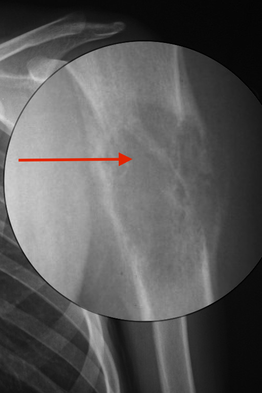
Onion skin ewing.jpeg| '''Onion skin sign ''': Also known as "multilayered periosteal reaction" demonstrates multiple concentric parallel layers of new bone adjacent to the cortex, reminiscent of the layers on an onion. Associated with osteosarcoma,Ewing sarcoma, and acute osteomyelitis (*) Parallel layers of new bone<br>[http://radiopaedia.org/articles/multilayered-periosteal-reaction<font size="-2">''Adapted from Radiopedia''</font>] | |||
Cockade_sign_bone_lipoma.jpeg |'''Cockade sign''': Classic appearance of an intraosseous lipoma of the calcaneus which presents as a well-defined lytic lesion with a central calcification resembling a cockade<br>[http://radiopaedia.org/articles/cockade-sign<font size="-2">''Adapted from Radiopedia''</font>] | |||
Geoskull multiple m.jpeg|'''Geographic skull sign''': radiographic appearance which is seen at eosinophilic granuloma. Destructive lytic bone lesion, edges of which may be bevelled, scalloped or confluent <br>[http://radiopaedia.org/articles/geographic-skull<font size="-2">''Adapted from Radiopedia''</font>] | |||
</gallery> | |||
===CT=== | |||
<gallery widths=200px> | |||
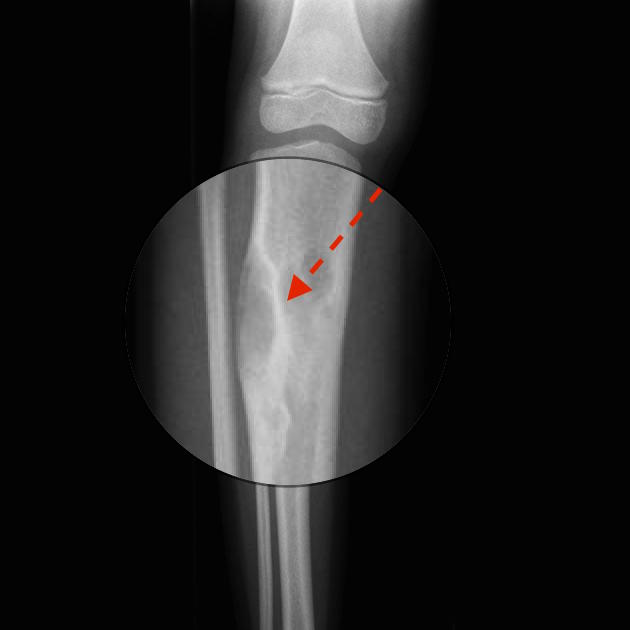
String sing ct osteosarcoma.jpeg | '''String sign ''': appearance a radiolucent cleavage plane between portions of the tumor and cortex of the affected bone<br>[http://radiopaedia.org/articles/string-sign-of-parosteal-osteosarcoma <font size="-2">''Adapted from Radiopedia''</font>] | |||
Osteoblastoma CT.jpeg |'''Osteoblastoma''': Internal matrix mineralisation is better appreciated on CT <br>[http://radiopaedia.org/articles/osteoblastoma<font size="-2">''Adapted from Radiopedia''</font>] | |||
</gallery> | |||
===MRI=== | |||
<gallery widths=200px> | |||
ONIONSIGN MRI.jpg | '''MRI- Onion skin sign '''<br>[http://radiopaedia.org/articles/multilayered-periosteal-reaction <font size="-2">''Adapted from Radiopedia''</font>] | |||
Enchondroma MRI.jpeg|'''MRI-Enchondroma''' : Well circumscribed somewhat lobulated masses replacing marrow<br>[http://radiopaedia.org/articles/multilayered-periosteal-reaction <font size="-2">''Adapted from Radiopedia''</font>] | |||
</gallery> | </gallery> | ||
Revision as of 17:26, 12 February 2016
|
Bone or Cartilage Mass Microchapters |
|
Diagnosis |
|---|
|
Case Studies |
|
Bone or cartilage mass imaging On the Web |
|
American Roentgen Ray Society Images of Bone or cartilage mass imaging |
|
Risk calculators and risk factors for Bone or cartilage mass imaging |
Editor-In-Chief: C. Michael Gibson, M.S., M.D. [1]Associate Editor(s)-in-Chief: Maria Fernanda Villarreal, M.D. [2]
Overview
Imaging
Plain Radiograph
CT
MRI
Gallery
Plain Radiograph
-
Osteosarcoma with Codman triangles: the tumor is essentially lytic and destructive with irregular, permeative margins, and soft tissue extension. Codman triangles (reactive periosteal new bone formation around the edges) are very prominent
Adapted from Creative Commons 3.0 -
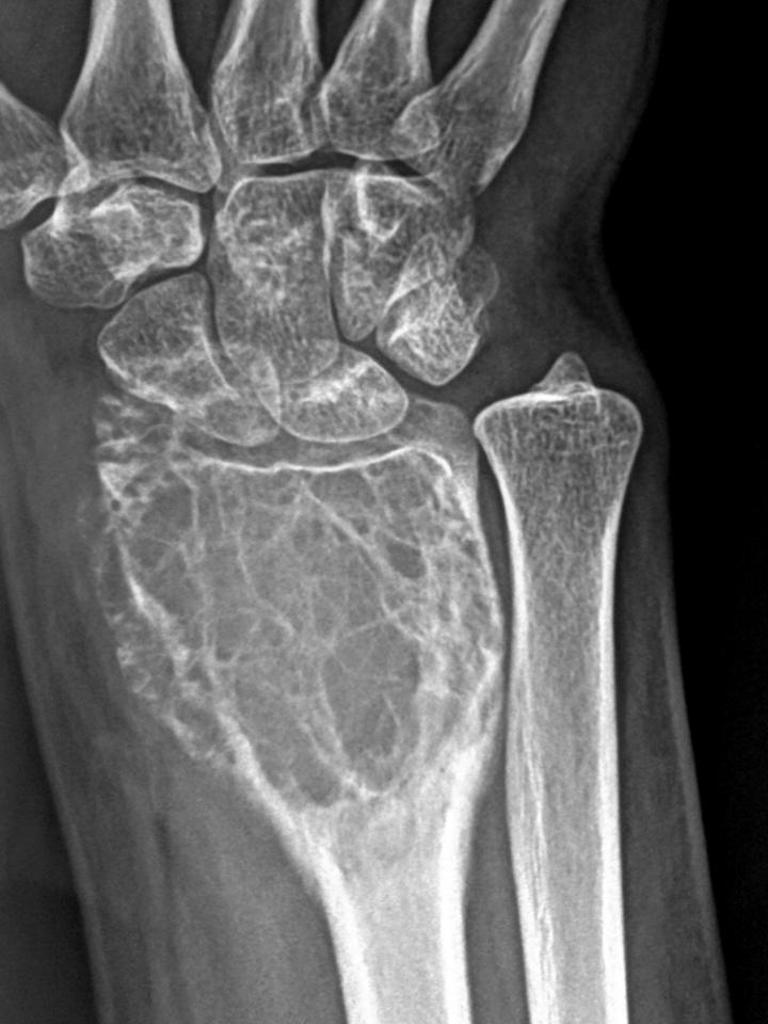
Giant cell tumor: located on distal radius
Adapted from Radiopedia -
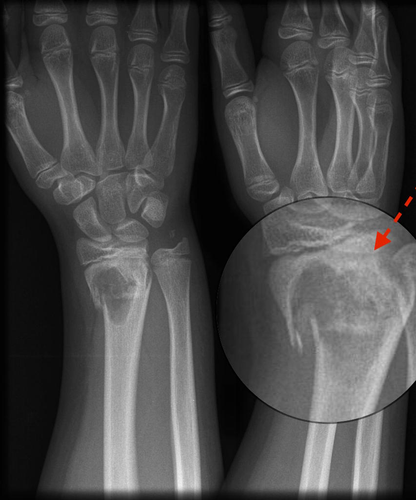
Pathological fracture: located in the metaphyseal region
Adapted from Radiopedia -
Rind sign: a lesion surrounded by a layer of thick, sclerotic reactive bone (rind) and is suggestive of fibrous dysplasia
Adapted from Radiopedia -
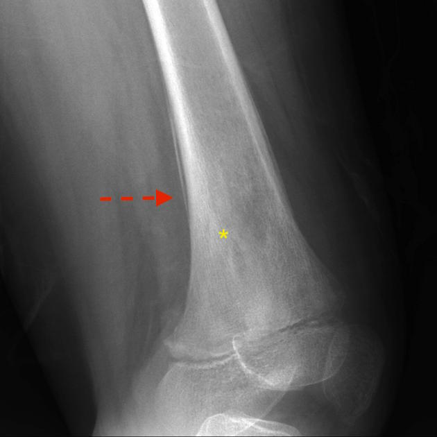
Sunburst appearance: a type of periosteal reaction giving the appearance of a sunburst secondary to an aggressive periostitis. Present in aggresive tumors, such as: osteosarcoma, Ewing sarcoma, and osteoblastic metastases (e.g. prostate, lung or breast cancer)
Adapted from Radiopedia -
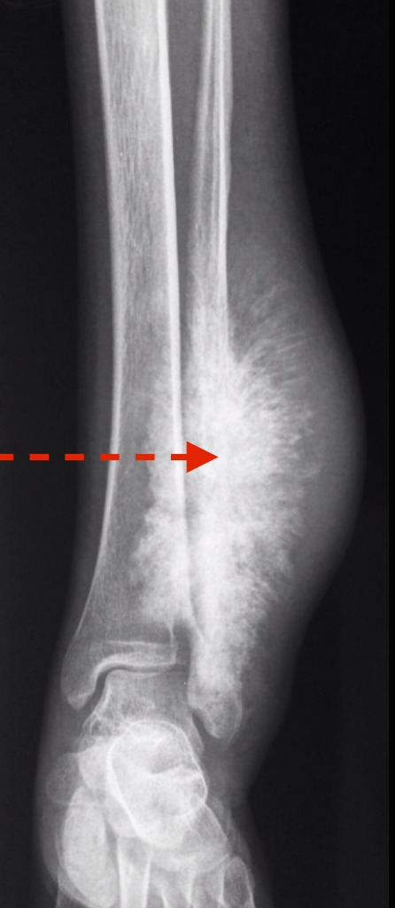
Onion skin sign : Also known as "multilayered periosteal reaction" demonstrates multiple concentric parallel layers of new bone adjacent to the cortex, reminiscent of the layers on an onion. Associated with osteosarcoma,Ewing sarcoma, and acute osteomyelitis (*) Parallel layers of new bone
Adapted from Radiopedia -
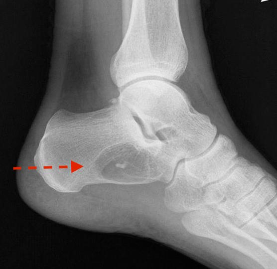
Cockade sign: Classic appearance of an intraosseous lipoma of the calcaneus which presents as a well-defined lytic lesion with a central calcification resembling a cockade
Adapted from Radiopedia -
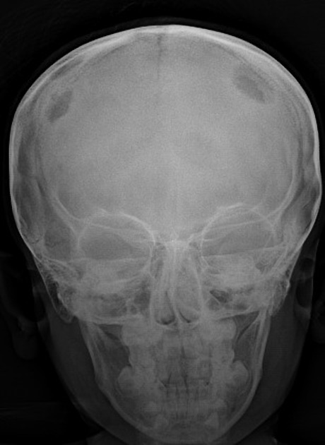
Geographic skull sign: radiographic appearance which is seen at eosinophilic granuloma. Destructive lytic bone lesion, edges of which may be bevelled, scalloped or confluent
Adapted from Radiopedia


CT
-
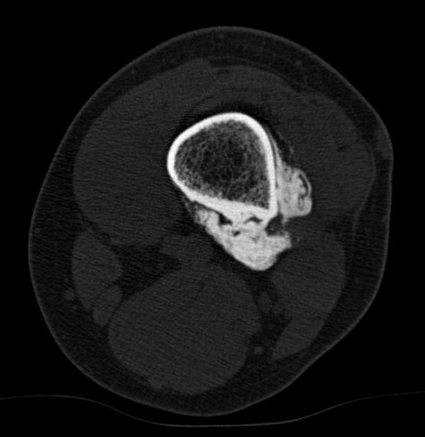
String sign : appearance a radiolucent cleavage plane between portions of the tumor and cortex of the affected bone
Adapted from Radiopedia -
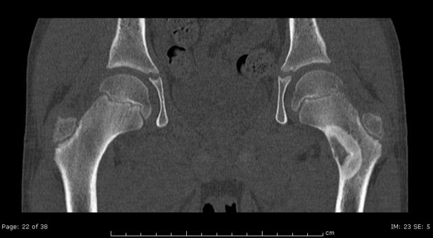
Osteoblastoma: Internal matrix mineralisation is better appreciated on CT
Adapted from Radiopedia


MRI
-
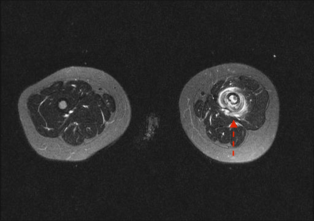
MRI- Onion skin sign
Adapted from Radiopedia -
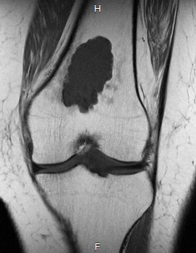
MRI-Enchondroma : Well circumscribed somewhat lobulated masses replacing marrow
Adapted from Radiopedia

