Rhabdomyosarcoma CT: Difference between revisions
Jump to navigation
Jump to search
No edit summary |
No edit summary |
||
| Line 8: | Line 8: | ||
*Some enhancement with contrast. | *Some enhancement with contrast. | ||
*Adjacent bony destruction seen in over 20% of cases. | *Adjacent bony destruction seen in over 20% of cases. | ||
===Rhabdomyosarcoma of biliary tract=== | |||
*CT may show a heterogenous or hypo-attenuating mass with [[Bile duct|biliary ductal dilatation]]. | |||
===Cardiac rhabdomyosarcoma=== | |||
*CT may show a smooth or irregular low-attenuation mass in a cardiac chamber. | |||
<div align="left"> | <div align="left"> | ||
| Line 16: | Line 22: | ||
</gallery> | </gallery> | ||
</div> | </div> | ||
==References== | ==References== | ||
Revision as of 13:58, 4 September 2015
|
Rhabdomyosarcoma Microchapters |
|
Diagnosis |
|---|
|
Treatment |
|
Case Studies |
|
Rhabdomyosarcoma CT On the Web |
|
American Roentgen Ray Society Images of Rhabdomyosarcoma CT |
Editor-In-Chief: C. Michael Gibson, M.S., M.D. [1]
Overview
CT
On CT scan, rhabdosarocma is characterized by:
- Soft tissue density.
- Some enhancement with contrast.
- Adjacent bony destruction seen in over 20% of cases.
Rhabdomyosarcoma of biliary tract
- CT may show a heterogenous or hypo-attenuating mass with biliary ductal dilatation.
Cardiac rhabdomyosarcoma
- CT may show a smooth or irregular low-attenuation mass in a cardiac chamber.
-
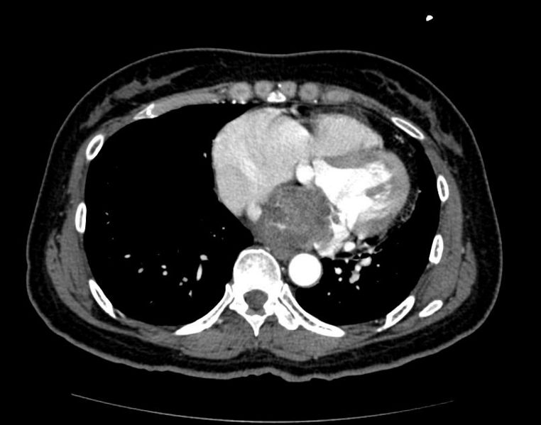
Cardiac Rhabdomyosarcoma Image courtesy of RadsWiki and copylefted
-
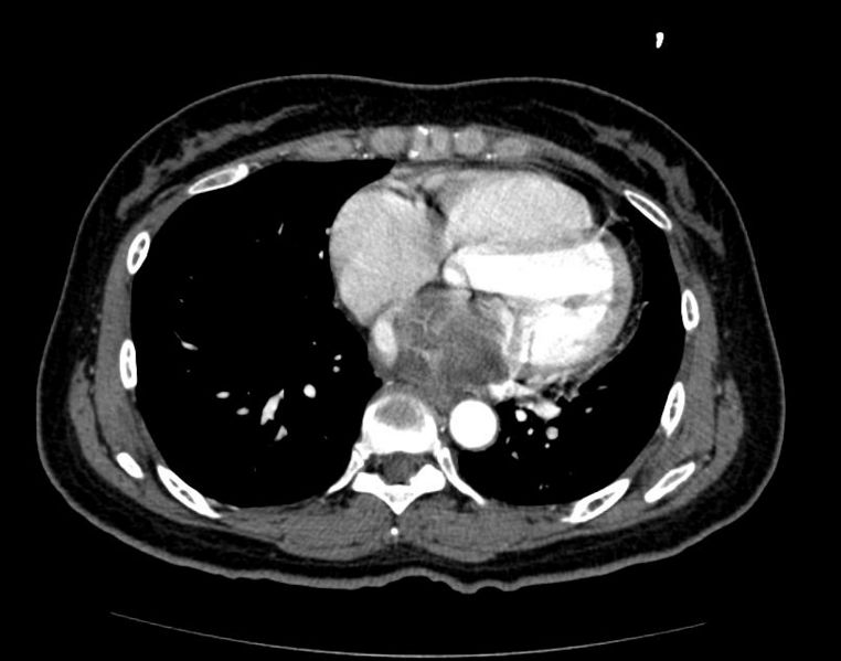
Cardiac Rhabdomyosarcoma Image courtesy of RadsWiki and copylefted
-
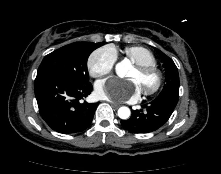
Cardiac Rhabdomyosarcoma Image courtesy of RadsWiki and copylefted


