Yersinia pestis infection physical examination
|
Yersinia pestis infection Microchapters |
|
Differentiating Yersinia Pestis Infection from other Diseases |
|---|
|
Diagnosis |
|
Treatment |
|
Case Studies |
|
Yersinia pestis infection physical examination On the Web |
|
American Roentgen Ray Society Images of Yersinia pestis infection physical examination |
|
Risk calculators and risk factors for Yersinia pestis infection physical examination |
Editor-In-Chief: C. Michael Gibson, M.S., M.D. [1] Associate Editor(s)-In-Chief: Yazan Daaboul; Serge Korjian
Overview
Apart from the presence of buboes, which are tender lymph nodes in patients infected with bubonic plague, the physical examination findings are not specific to plague. Nonetheless, physical examination is crucial to evaluate for the presence of target organ damage or the progression and worsening of infection burden in these patients.[1]
Physical Examination
Buboes, which are fixed tender lymphadenopathy in patients with bubonic plague, are characteristic findings on physical examination.[1] Otherwise, patients with plague infection generally have non-specific physical examination findings. Nonetheless, physical examination is key in all cases of plague for the evaluation of the patient's clinical picture and monitoring for clinical response.[1]
General Appearance
Patients infected with plague are generally toxic-looking.
Vital Signs
- Fever is invariably present in all patients
- Tachycardia
- Tachypnea
- Hypotension in septicemic plague
HEENT
- Pharyngeal erythema
- Tonsilar enlargement
- Meningismus in plague complicated by meningitis
Skin
- Papules, pustules, ulcers, or vesicles surrounded by local erythema at site of infection
- Fixed tender lymphadenopathy of 1-10 cm in diameter in bubonic plague that may be present anywhere: axillary, cervical, postauricular, inguinal, epitrochlear, or popliteal. Lymph nodes may demonstrate fluctuance, suppuration, and may eventually drain.
-
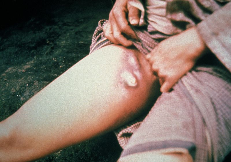 Inguinal lymphadenoapthy
 -
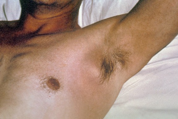 Axillary lymphadenopathy


Chest
- Auscultation may be minimal despite worsening infectious burden
- Dullness to percussion due to segmental pulmonary consolidation
- Decreased breath sounds over affected area
Abdomen
- Intra-abdominal buboes may present with abdominal tenderness, guarding, or signs of peritoneal irritation, such as rebound tenderness and abdominal rigidity.
- Hepatomegaly
Extremities
- Black gangrenous distal upper and lower extremities (shown below)
-
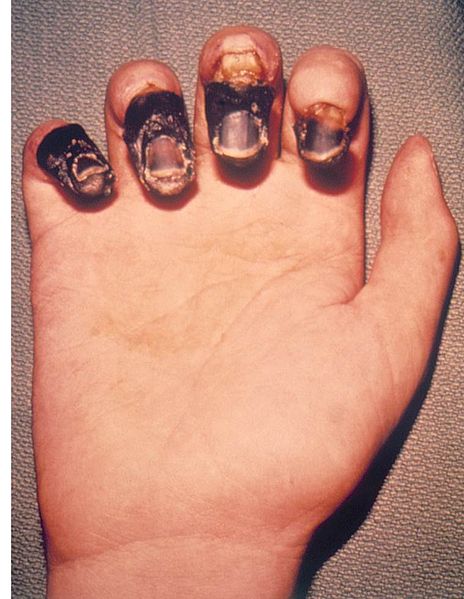 Acral ganagre of the digits in bubonic plague
 -
![Dorsal view of a 59 year-old man’s right hand who had been infected by the plague bacterium, Yersinia pestis, after having come into contact with both an infected cat, and a dead mouse in his neighborhood. Adapted from Public Health Image Library (PHIL), Centers for Disease Control and Prevention.[2]](/images/b/b6/Plague1.jpg) Dorsal view of a 59 year-old man’s right hand who had been infected by the plague bacterium, Yersinia pestis, after having come into contact with both an infected cat, and a dead mouse in his neighborhood. Adapted from Public Health Image Library (PHIL), Centers for Disease Control and Prevention.[2]
Dorsal view of a 59 year-old man’s right hand who had been infected by the plague bacterium, Yersinia pestis, after having come into contact with both an infected cat, and a dead mouse in his neighborhood. Adapted from Public Health Image Library (PHIL), Centers for Disease Control and Prevention.[2] -
![Dorsal view of a 59 year-old man’s hands who had been infected by the plague bacterium, Yersinia pestis, after having come into contact with both an infected cat, and a dead mouse in his neighborhood. Adapted from Public Health Image Library (PHIL), Centers for Disease Control and Prevention.[2]](/images/3/30/Plague2.jpg) Dorsal view of a 59 year-old man’s hands who had been infected by the plague bacterium, Yersinia pestis, after having come into contact with both an infected cat, and a dead mouse in his neighborhood. Adapted from Public Health Image Library (PHIL), Centers for Disease Control and Prevention.[2]
Dorsal view of a 59 year-old man’s hands who had been infected by the plague bacterium, Yersinia pestis, after having come into contact with both an infected cat, and a dead mouse in his neighborhood. Adapted from Public Health Image Library (PHIL), Centers for Disease Control and Prevention.[2] -
![Palmar view of a 59 year-old man’s right hand who had been infected by the plague bacterium, Yersinia pestis, after having come into contact with both an infected cat, and a dead mouse in his neighborhood. Adapted from Public Health Image Library (PHIL), Centers for Disease Control and Prevention.[2]](/images/c/c9/Plague3.jpg) Palmar view of a 59 year-old man’s right hand who had been infected by the plague bacterium, Yersinia pestis, after having come into contact with both an infected cat, and a dead mouse in his neighborhood. Adapted from Public Health Image Library (PHIL), Centers for Disease Control and Prevention.[2]
Palmar view of a 59 year-old man’s right hand who had been infected by the plague bacterium, Yersinia pestis, after having come into contact with both an infected cat, and a dead mouse in his neighborhood. Adapted from Public Health Image Library (PHIL), Centers for Disease Control and Prevention.[2] -
![Dorsal view of a 59 year-old man’s feet who had been infected by the plague bacterium, Yersinia pestis, after having come into contact with both an infected cat, and a dead mouse in his neighborhood. Adapted from Public Health Image Library (PHIL), Centers for Disease Control and Prevention.[2]](/images/d/d5/Plague4.jpg) Dorsal view of a 59 year-old man’s feet who had been infected by the plague bacterium, Yersinia pestis, after having come into contact with both an infected cat, and a dead mouse in his neighborhood. Adapted from Public Health Image Library (PHIL), Centers for Disease Control and Prevention.[2]
Dorsal view of a 59 year-old man’s feet who had been infected by the plague bacterium, Yersinia pestis, after having come into contact with both an infected cat, and a dead mouse in his neighborhood. Adapted from Public Health Image Library (PHIL), Centers for Disease Control and Prevention.[2]

![Dorsal view of a 59 year-old man’s right hand who had been infected by the plague bacterium, Yersinia pestis, after having come into contact with both an infected cat, and a dead mouse in his neighborhood. Adapted from Public Health Image Library (PHIL), Centers for Disease Control and Prevention.[2]](/index.php/File:Plague1.jpg)
![Dorsal view of a 59 year-old man’s hands who had been infected by the plague bacterium, Yersinia pestis, after having come into contact with both an infected cat, and a dead mouse in his neighborhood. Adapted from Public Health Image Library (PHIL), Centers for Disease Control and Prevention.[2]](/index.php/File:Plague2.jpg)
![Palmar view of a 59 year-old man’s right hand who had been infected by the plague bacterium, Yersinia pestis, after having come into contact with both an infected cat, and a dead mouse in his neighborhood. Adapted from Public Health Image Library (PHIL), Centers for Disease Control and Prevention.[2]](/index.php/File:Plague3.jpg)
![Dorsal view of a 59 year-old man’s feet who had been infected by the plague bacterium, Yersinia pestis, after having come into contact with both an infected cat, and a dead mouse in his neighborhood. Adapted from Public Health Image Library (PHIL), Centers for Disease Control and Prevention.[2]](/index.php/File:Plague4.jpg)
- Warm skin in septic patients
- Petechiae in patients with disseminated intravascular coagulopathy (DIC)