Vertebrate trachea
Editor-In-Chief: C. Michael Gibson, M.S., M.D. [1]
Overview
The tracheartes, or windpipe, is a tube that has an inner diameter of about 20-25 mm and a length of about 10-16cm. It commences at the larynx(at the level vertebral level of C6 in humans) and bifurcates into the primary (main) bronchi (at the vertebral level of T4/T5 in humans) in mammals, and from the pharynx to the syrinx in birds, allowing the passage of air to the lungs. It is lined with pseudostratified ciliated columnar epithelium cells with mucosae goblet cells which produce mucus. This lines the cells of the trachea to trap inhaled foreign particles which the cilia then waft upwards towards their larynx and then the pharynx where it can then be swallowed into the stomach.
In humans there are about 15 – 20 incomplete C-shaped cartilaginous rings which reinforces the anterior and lateral sides of the trachea to protect and maintain the airway open. There is a piece of smooth muscle connecting the ends off the incomplete cartilaginous rings called the Trachealis muscle. This contracts reducing the size of the lumen of the trachea to increase the air flow rate during coughing. The esophagus lies posteriorly to the trachea. The cartilaginous rings are incomplete because this allows the trachea to collapse slightly to allow food to pass down the esophagus. The epiglottis is the flap that closes the trachea during swallowing to prevent swallowed matter from entering the trachea.
Clinical significance
Endotracheal intubation is the medical procedure of inserting an artificial tube into the trachea to provide a secure route for ventilating the lungs.
Tracheotomy is a surgical procedure of making an opening in the front of the neck that extends to the lumen of the trachea, a short tube called a tracheostomy tube is inserted through this opening, entering below the level of the larynx and vocal cords.
Additional images
-
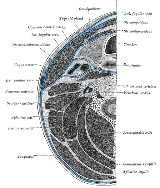
Section of the neck at about the level of the sixth cervical vertebra.
-
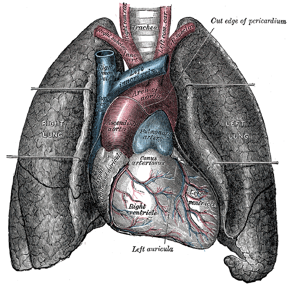
Front view of heart and lungs.
-
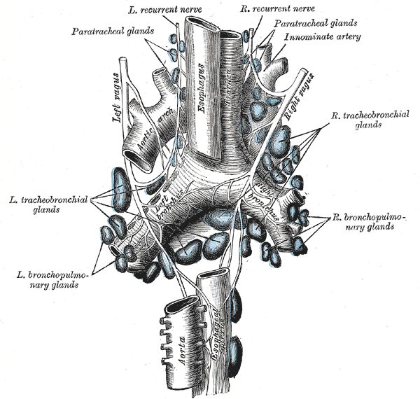
The tracheobronchial lymph glands.
-
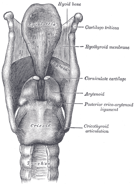
Ligaments of the larynx. Posterior view.
-
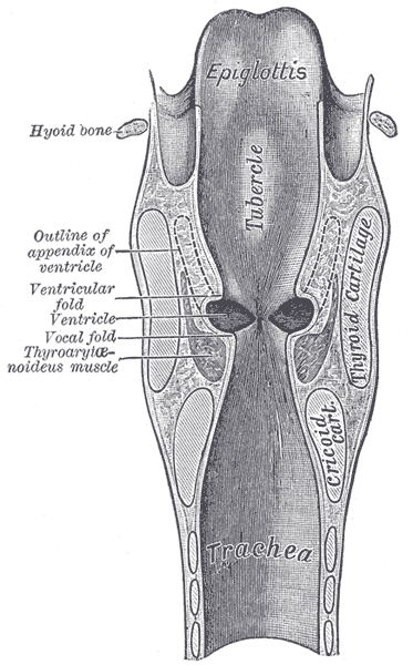
Coronal section of larynx and upper part of trachea.
-
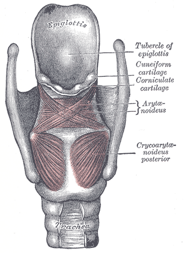
Muscles of larynx. Posterior view.
-
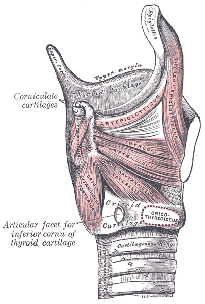
Muscles of larynx. Side view. Right lamina of thyroid cartilage removed.
-
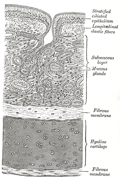
Transverse section of trachea.
-
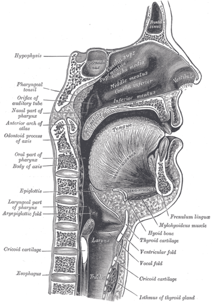
Sagittal section of nose mouth, pharynx, and larynx.
-
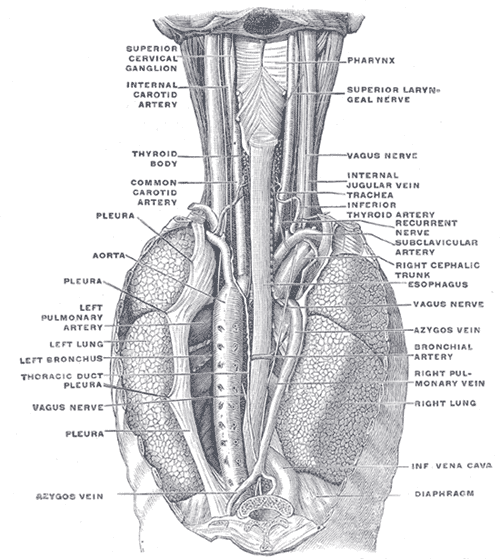
The position and relation of the esophagus in the cervical region and in the posterior mediastinum. Seen from behind.
-
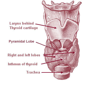
Thyroid
-
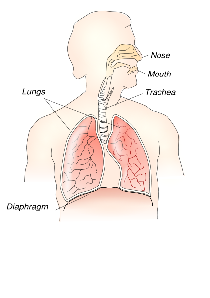
Respiratory system
-
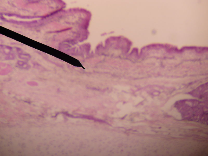
Microscopic cross section of human trachea.


Template:Respiratory system Template:Lung
ar:قصبة هوائية ca:Tràquea cs:Průdušnice de:Luftröhre et:Hingetoru eo:Traĥeo eu:Trakea hr:Dušnik id:Trakea it:Trachea he:קנה הנשימה la:Trachea lt:Trachėja mk:Дишник nl:Luchtpijp no:Luftrør sr:Душник sh:Dušnik fi:Henkitorvi sv:Luftstrupe uk:Трахея yi:ווינט רער