Sacrum
|
WikiDoc Resources for Sacrum |
|
Articles |
|---|
|
Media |
|
Evidence Based Medicine |
|
Clinical Trials |
|
Ongoing Trials on Sacrum at Clinical Trials.gov Clinical Trials on Sacrum at Google
|
|
Guidelines / Policies / Govt |
|
US National Guidelines Clearinghouse on Sacrum
|
|
Books |
|
News |
|
Commentary |
|
Definitions |
|
Patient Resources / Community |
|
Directions to Hospitals Treating Sacrum Risk calculators and risk factors for Sacrum
|
|
Healthcare Provider Resources |
|
Continuing Medical Education (CME) |
|
International |
|
|
|
Business |
|
Experimental / Informatics |
Editor-In-Chief: C. Michael Gibson, M.S., M.D. [1]
Overview
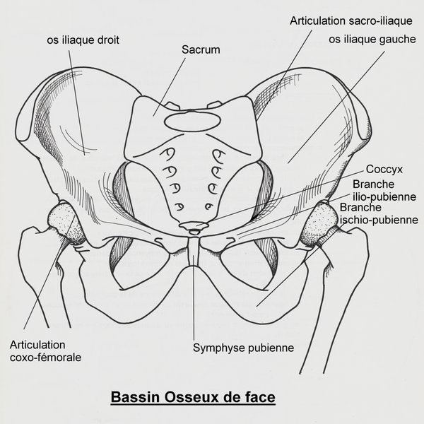
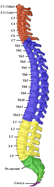
The sacrum is a large, triangular bone at the base of the spine and at the upper and back part of the pelvic cavity, where it is inserted like a wedge between the two hip bones. Its upper part connects with the last lumbar vertebra, and bottom part with the coccyx (tailbone).
It is curved upon itself and placed obliquely (that is, tilted forward). It is concave facing forwards, thus its curvature is considered a kyphosis. The base projects forward as the sacral promontory internally, and articulates with the last lumbar vertebra to form the prominent sacrovertebral angle. The central part is curved outward towards the posterior, allowing greater room for the pelvic cavity.
Etymology
The name is derived from the Latin sacer, "sacred", a translation of the Greek hieron (osteon), meaning sacred or strong bone.[1] This is supposedly derived from the belief that it could not be destroyed and was the part that would allow rising from the dead.[2]
Parts
- The pelvic surface of sacrum is concave from above downward, and slightly so from side to side.
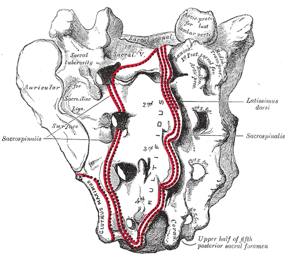
- The dorsal surface of sacrum is convex and narrower than the pelvic.
- The lateral surface of sacrum is broad above, but narrowed into a thin edge below.
- The base of the sacrum, which is broad and expanded, is directed upward and forward.
- The apex (apex oss. sacri) is directed downward, and presents an oval facet for articulation with the coccyx.
- The vertebral canal (canalis sacralis; sacral canal) runs throughout the greater part of the bone; above, it is triangular in form; below, its posterior wall is incomplete, from the non-development of the laminae and spinous processes. It lodges the sacral nerves, and its walls are perforated by the anterior and posterior sacral foramina through which these nerves pass out.
Articulations
The sacrum articulates with four bones:
- the last lumbar vertebra above
- the coccyx below
- the hip bone on either side
Although in most people the sacro-iliac joints are tightly bound and immobile, some are able to rotate the sacrum forward a few degrees vis-à-vis the ilia. This motion is sometimes called "nutation", and the reverse motion "counter-nutation."[3]
It is called the sacrum when referred to all of the parts combined, but sacral vertebrae when referred individually.
Sexual dimorphism
The sacrum is noticeably sexually dimorphic (differently-shaped in males and females).
In the female the sacrum is shorter and wider than in the male; the lower half forms a greater angle with the upper; the upper half is nearly straight, the lower half presenting the greatest amount of curvature. The bone is also directed more obliquely backward; this increases the size of the pelvic cavity and renders the sacrovertebral angle more prominent.
In the male the curvature is more evenly distributed over the whole length of the bone, and is altogether greater than in the female.
Variations
The sacrum, in some cases, consists of six pieces [2]; occasionally the number is reduced to four [3]. The bodies of the first and second vertebrae may fail to unite.
Sometimes the uppermost transverse tubercles are not joined to the rest of the ala on one or both sides, or the sacral canal may be open throughout a considerable part of its length, in consequence of the imperfect development of the laminae and spinous processes.
The sacrum, also, varies considerably with respect to its degree of curvature
Additional images
-
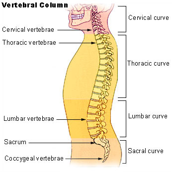
Vertebral column.
-
-
Sacrum, dorsal surface.
-
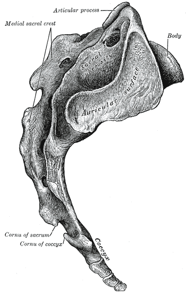
Lateral surfaces of sacrum and coccyx.
-
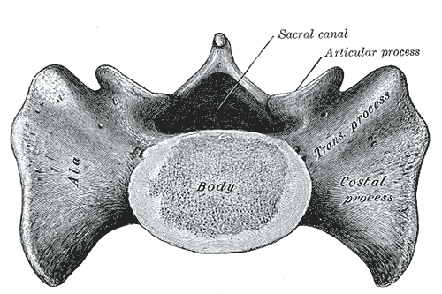
Base of sacrum.
-
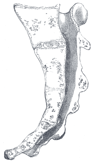
Median sagittal section of the sacrum.
-
Vertebral column.
-
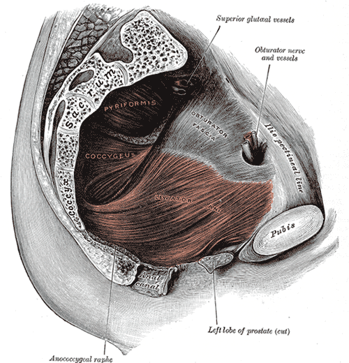
Left Levator ani from within.
-
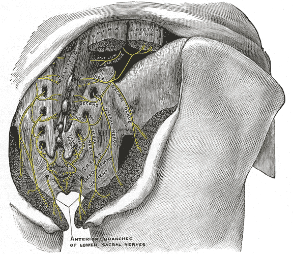
The posterior divisions of the sacral nerves.
-
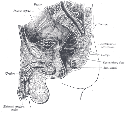
Median sagittal section of male pelvis.
-
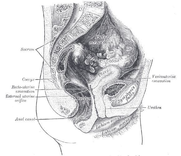
Median sagittal section of female pelvis.


See also
External links
- Template:SUNYAnatomyLabs - "The Female Pelvis: Articulated bones of pelvis"
- Template:SUNYAnatomyLabs - "The Female Pelvis: Bones"
- Template:EMedicineDictionary
References
Template:Gray's Template:Spine
ca:Sacre de:Kreuzbein eo:Sakro (anatomio) it:Osso sacro he:סקרום la:Os sacrum lt:Kryžkaulis nl:Heiligbeen sk:Krížová kosť fi:Ristiluu sv:Korsben uk:Крижова кістка