Renovascular disease other imaging findings
Jump to navigation
Jump to search
|
Renovascular disease Microchapters |
|
Diagnosis |
|---|
|
Treatment |
|
Case Studies |
|
Renovascular disease other imaging findings On the Web |
|
American Roentgen Ray Society Images of Renovascular disease other imaging findings |
|
Risk calculators and risk factors for Renovascular disease other imaging findings |
Editor-In-Chief: C. Michael Gibson, M.S., M.D. [1]
Please help WikiDoc by adding more content here. It's easy! Click here to learn about editing.
Overview
Other Imaging Studies
Diagnostic Methods to Detect Renal Artery Stenosis - ACC/AHA Guidelines (DO NOT EDIT)
| “ |
CLASS I
CLASS III
|
” |
Renal Arteriography
Renal Arteriography
- Abdominal Aortogram: identification of ostia of the renal arteries and accessory renal arteries (25% of population)
- Arteriography should include both the arterial phase and the nephrographic phase
- Disease involving renal bifurcations require cranial or caudal angulation to open out the lesion
- Evidence of aortic atheroma: technique of no-touch angiography is recommended
-
 Renal artery stenosis
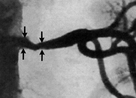
 -
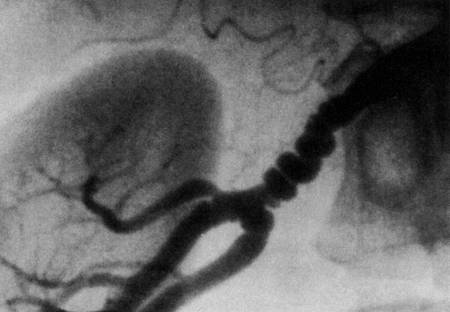 Fibromscular dysplasia

