Pulmonary artery
Editor-In-Chief: C. Michael Gibson, M.S., M.D. [1]
Please Take Over This Page and Apply to be Editor-In-Chief for this topic: There can be one or more than one Editor-In-Chief. You may also apply to be an Associate Editor-In-Chief of one of the subtopics below. Please mail us [2] to indicate your interest in serving either as an Editor-In-Chief of the entire topic or as an Associate Editor-In-Chief for a subtopic. Please be sure to attach your CV and or biographical sketch.
The pulmonary arteries carry blood from the heart to the lungs. They are the only arteries (other than umbilical arteries in the fetus) that carry deoxygenated blood.
In the human heart, the pulmonary trunk (pulmonary artery or main pulmonary artery) begins at the base of the right ventricle. It is short and wide - approximately 5 cm (2 inches) in length and 3 cm (1.2 inches) in diameter. It then branches into two pulmonary arteries (left and right), which deliver deoxygenated blood to the corresponding lung.
Role in disease
Pulmonary hypertension occurs alone and as a consequence of a number of lung diseases. It can be a consequence of heart disease (Eisenmenger's syndrome) but equally a cause (right-ventricular heart failure); it also occurs as a consequence of pulmonary embolism and scleroderma. It is characterised by reduced exercise tolerance. Severe forms, generally, have a dismal prognosis.
Additional images
-
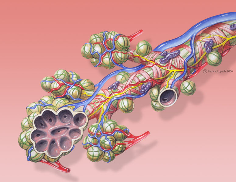
Bronchial anatomy
-
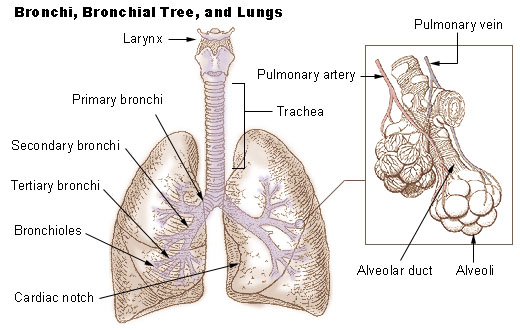
Bronchi, bronchial tree, and lungs
-
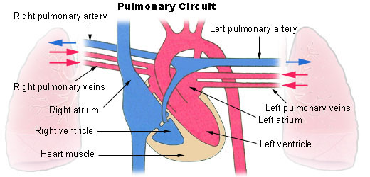
Pulmonary circuit
-
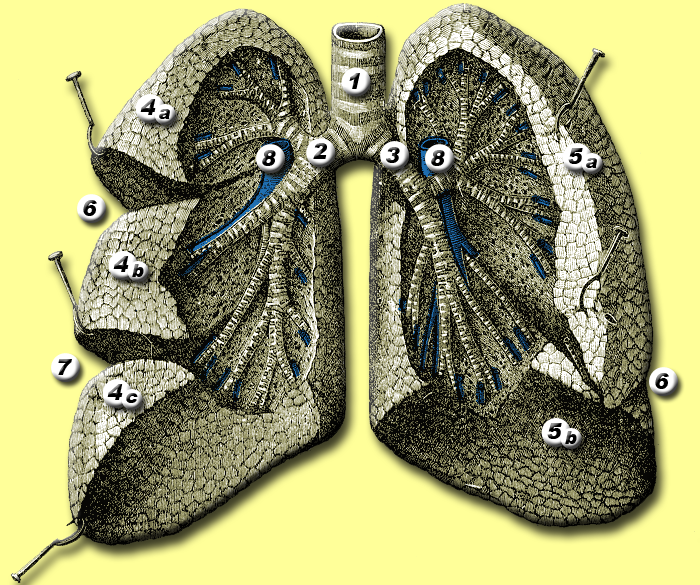
Anatomy of lungs.
-
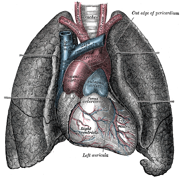
Front view of heart and lungs.
-
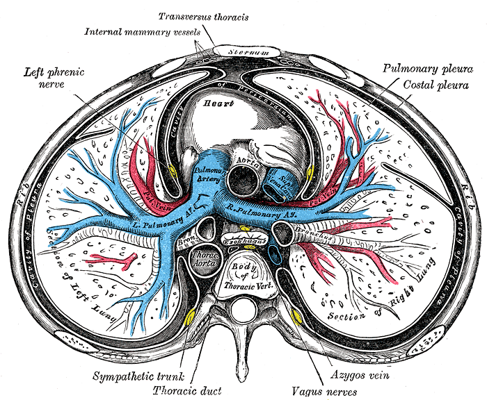
Transverse section of thorax, showing relations of pulmonary artery.
-

Base and diaphragmatic surface of heart.
-
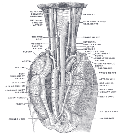
The position and relation of the esophagus in the cervical region and in the posterior mediastinum. Seen from behind.


See also
- Chronic obstructive lung disease
- Pulmonary hypertension
- Thromboembolic disease
- Pulmonary circulation
- Rasmussen's aneurysm
External links
- Template:GPnotebook
- Template:EMedicineDictionary
- Template:SUNYAnatomyLabs - "Heart: The Pericardial sac and Great vessels"
- Template:SUNYAnatomyLabs - "Heart: Openings of Great Vessels into the Pericardial Sac"
- Template:SUNYAnatomyFigs - "Mediastinal surface of the right lung."
- Template:SUNYAnatomyFigs - "Mediastinal surface of the left lung."
- Histology image: 13802loa – Histology Learning System at Boston University
Template:Arteries of chest Template:SIB
ar:الشريان الرئوي gl:Arteria pulmonar it:Arteria polmonare nl:Longslagader nn:Lungepulsåre simple:Pulmonary artery sr:Плућно артеријско стабло