Papilledema physical examination
|
Papilledema |
|
Diagnosis |
|---|
|
Treatment |
|
Case Studies |
|
Papilledema physical examination On the Web |
|
American Roentgen Ray Society Images of Papilledema physical examination |
|
Risk calculators and risk factors for Papilledema physical examination |
Please help WikiDoc by adding more content here. It's easy! Click here to learn about editing.
Editor-In-Chief: C. Michael Gibson, M.S., M.D. [1] Associate Editor(s)-in-Chief: Kalsang Dolma, M.B.B.S.[2]
Overview
The signs of papilledema include blurring of the margins of the optic disc, edema, and hemorrhages on fundoscopy.
Physical Examination
Vitals
Look for blood pressure to rule out hypertension
Eyes
-
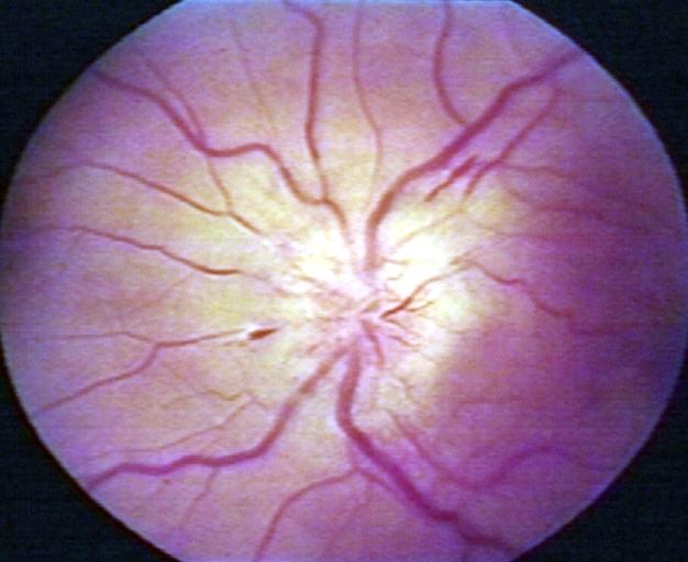 Papilledema.
 -
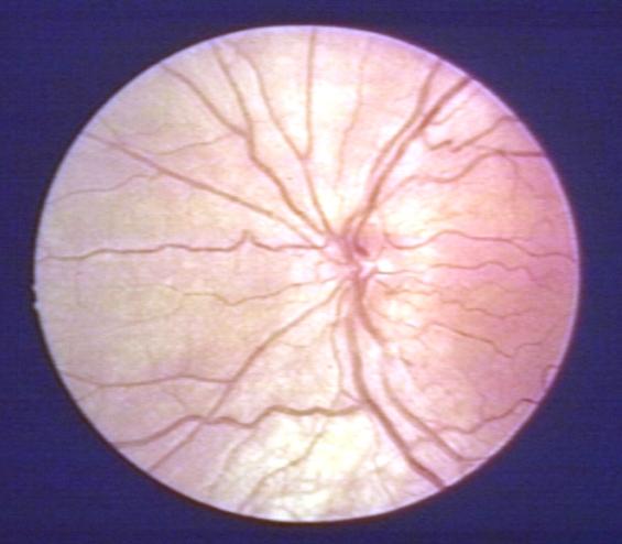 Pseudopapilledema.


Checking the eyes for signs of papilledema should be carried out whenever there is a clinical suspicion of raised intracranial pressure. Because of the (rare) possibility of a brain tumor, pseudotumor cerebri or cerebral venous sinus thrombosis, all of which can increase intracranial pressure, this examination has become common for patients suffering from headaches.
There are 10 hallmarks of papilledema seen by using opthalmoscope:
- Blurring of the disc margins
- Filling in of the optic disc cup
- Anterior bulging of the nerve head
- Edema of the nerve fiber layer
- Retinal or choroidal folds
- Congestion of retinal veins
- Peripapillary hemorrhages
- Hyperemia of the optic nerve head
- Nerve fiber layer infarcts
- Hard exudates of the optic disc
Frisén has proposed a useful staging scheme for papilledema with good sensitivity and specificity based on the ophthalmoscopic signs of disturbed axoplasmic transport. It has been modified recently with a key finding added for each stage or grade.
- Grade 0 Represents a normal optic disc.
- Grade 1 Presence of a C-shaped or reverse C-shaped halo of peripapillary edema obscuring the retina adjacent to the optic disc. The temporal border of the optic disc is spared presumably due to the fine caliber of these axons.
- Grade 2 The C-shaped halo becomes circumferential with grade 2 papilledema.
- Grade 3 Complete obscuration of at least one major vessel as it leaves the optic disc.
- Grade 4 Complete obscuration of at least one major vessel on the optic disc.
- Grade 5 Total obscuration of at least one vessel on the disc and leaving the disc and at least partial obscuration of all major vessels leaving or on the disc.
On visual field examination, the physician may elicit an enlarged blind spot; the visual acuity may remain relatively intact until papilledema is severe or prolonged.