Mucoepidermoid carcinoma physical examination
|
Mucoepidermoid carcinoma Microchapters |
|
Differentiating Mucoepidermoid Carcinoma from other Diseases |
|---|
|
Diagnosis |
|
Treatment |
|
Case Studies |
|
Mucoepidermoid carcinoma physical examination On the Web |
|
American Roentgen Ray Society Images of Mucoepidermoid carcinoma physical examination |
|
Risk calculators and risk factors for Mucoepidermoid carcinoma physical examination |
Editor-In-Chief: C. Michael Gibson, M.S., M.D. [1]Associate Editor(s)-in-Chief: Badria Munir M.B.B.S.[2] , Maria Fernanda Villarreal, M.D. [3]
Overview
Physical examination findings of mucoepidermoid carcinoma depend on the size and location of the tumor. Physical examination of patients with mucoepidermoid carcinoma is usually remarkable for tenderness and intraoral bluish-red mucosa.
Physical Examination
The following physical examination findings may be present among patients with mucoepidermoid carcinoma:[1][1]
Appearance
Vital Signs
- Fever may be present
HEENT
- Unilateral otorrhea
- Hearing acuity may be reduced
- Bluish-red intraoral mucosa
NECK
- Carotid bruits may be auscultated unilaterally using the bell/diaphragm of the otoscope
- Unilateral lymphadenopathy
Image Gallery
-
-
-
-
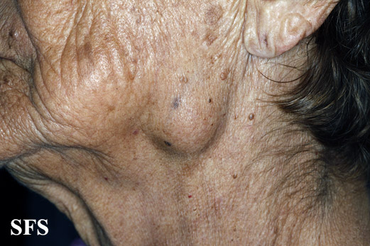
![Parotid swelling With permission from Dermatology Atlas.[2]](/images/a/aa/Parotitis01.jpg) Parotid swelling With permission from Dermatology Atlas.[2]
Parotid swelling With permission from Dermatology Atlas.[2] -
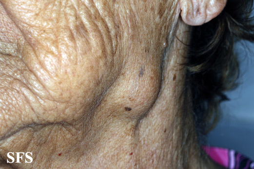
![Parotid swelling With permission from Dermatology Atlas.[2]](/images/f/f8/Parotitis02.jpg) Parotid swelling With permission from Dermatology Atlas.[2]
Parotid swelling With permission from Dermatology Atlas.[2] -
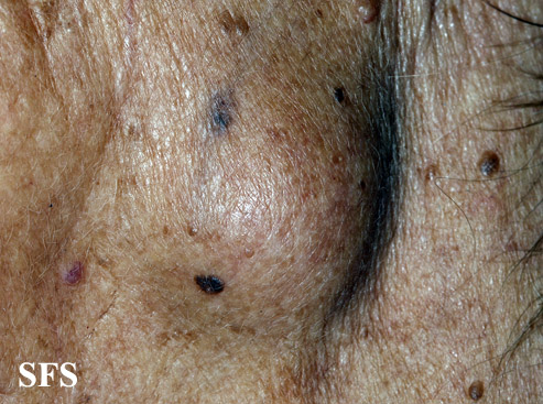
![Parotid swelling With permission from Dermatology Atlas.[2]](/images/3/3d/Parotitis03.jpg) Parotid swelling With permission from Dermatology Atlas.[2]
Parotid swelling With permission from Dermatology Atlas.[2]


![Parotid swelling With permission from Dermatology Atlas.[2]](/index.php/File:Parotitis01.jpg)
![Parotid swelling With permission from Dermatology Atlas.[2]](/index.php/File:Parotitis02.jpg)
![Parotid swelling With permission from Dermatology Atlas.[2]](/index.php/File:Parotitis03.jpg)
Video Gallery
{{#ev:youtube|ZpmE1y51IU8}}
{{#ev:youtube|Gx1vVwA5AFU}}
{{#ev:youtube|3lHRVAOJq3k}}
{{#ev:youtube|R-_rt857H5c}}