Leydig cell
|
WikiDoc Resources for Leydig cell |
|
Articles |
|---|
|
Most recent articles on Leydig cell Most cited articles on Leydig cell |
|
Media |
|
Powerpoint slides on Leydig cell |
|
Evidence Based Medicine |
|
Clinical Trials |
|
Ongoing Trials on Leydig cell at Clinical Trials.gov Clinical Trials on Leydig cell at Google
|
|
Guidelines / Policies / Govt |
|
US National Guidelines Clearinghouse on Leydig cell
|
|
Books |
|
News |
|
Commentary |
|
Definitions |
|
Patient Resources / Community |
|
Patient resources on Leydig cell Discussion groups on Leydig cell Patient Handouts on Leydig cell Directions to Hospitals Treating Leydig cell Risk calculators and risk factors for Leydig cell
|
|
Healthcare Provider Resources |
|
Causes & Risk Factors for Leydig cell |
|
Continuing Medical Education (CME) |
|
International |
|
|
|
Business |
|
Experimental / Informatics |
Leydig cells, also known as interstitial cells of Leydig, are found adjacent to the seminiferous tubules in the testicle. They can secrete testosterone and are often closely related to nerves. Leydig cells have round vesicular nuclei and a granular eosinophilic cytoplasm.
Nomenclature
Leydig cells are named after the German anatomist Franz Leydig, who discovered them in 1850.[1]
Functions
Leydig cells release a class of hormones called androgens (19-carbon steroids). They secrete testosterone, androstenedione and dehydroepiandrosterone (DHEA), when stimulated by the pituitary hormone luteinizing hormone (LH). LH increases cholesterol desmolase activity (an enzyme associated with the conversion of cholesterol to pregnenolone), leading to testosterone synthesis secretion by Leydig cells.
Follicle-stimulating hormone (FSH) increases the response of Leydig cells to LH by increasing the number of LH receptors expressed on Leydig cells.
Ultrastructure
Leydig cells are polygonal, eosinophilic cells with a round vesicular nucleus and contain lipid droplets. They contain abundant smooth endoplasmic reticulum, which accounts for their eosinophilia. Frequently, lipofuscin pigment and rod-shaped crystal-like structures (Reinke's crystals) are found.[2][3]
Development
Leydig cells form during the 16th and 20th week of gestation and are quiescent until puberty.
Additional images
-
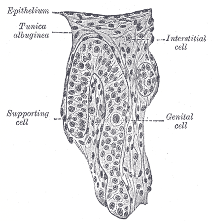
Section of a genital cord of the testis of a human embryo 3.5 cm. long.
References
- ↑ Template:WhoNamedIt
- ↑ Al-Agha O, Axiotis C (2007). "An in-depth look at Leydig cell tumor of the testis". Arch Pathol Lab Med. 131 (2): 311–7. PMID 17284120.
- ↑ Ramnani, Dharam M (2005-01-25). "Leydig Cell Tumor : Reinke's Crystalloids". Retrieved 2007-03-28.
See also
External links
- Histology image: 16907loa – Histology Learning System at Boston University
- Reproductive Physiology
- Diagram at umassmed.edu
Template:Male reproductive system
de:Leydig-Zelle id:Sel Leydig it:Cellula di Leydig he:תא ליידיג la:Cellulae Leydig nl:Leydigcel sk:Leydigova bunka
