Lens (anatomy)
Template:Infobox Anatomy Editor-In-Chief: C. Michael Gibson, M.S., M.D. [1]
Overview
The lens is a transparent, biconvex (lentil-shaped) structure in the eye that, along with the cornea, helps to refract light to be focused on the retina. The lens, by changing shape, functions to change the focal distance of the eye so that it can focus on objects at various distances, thus allowing a sharp image of the object of interest to be formed on the retina. This adjustment of the lens is known as accommodation (see also Accomodation, below). It is similar to the focusing of a photographic camera via movement of its lenses.
The lens is also known as the aquula (Latin, a little stream, dim. of aqua, water) or crystalline lens. In humans, the refractive power of the lens in its natural environment is approximately 18 dioptres, roughly one-third of the eye's total power.
Position, size, and shape
The lens is located in the anterior segment of the eye. Anterior to the lens is the iris, which regulates the amount of light entering the eye. The lens is suspended in place by the zonular fibers, which attach to the lens near its equatorial line and connect the lens to the ciliary body. Posterior to the lens is the vitreous body. The lens has an ellipsoid, biconvex shape. In the adult, the lens is typically 10 mm in diameter and has an axial length of 4 mm, though it is important to note that the size and shape can change due to accommodation and because the lens continues to grow throughout a person’s lifetime.[1]
Lens Structure and Function
The lens is comprised of three main parts: the lens capsule, the lens epithelium, and the lens fibers. The lens capsule forms the outermost layer of the lens and the lens fibers form the bulk of the interior of the lens. The cells of the lens epithelium, located between the lens capsule and the outermost layer of lens fibers, are found only on the anterior side of the lens.
Lens Capsule
The lens capsule is a smooth, transparent basement membrane that completely surrounds the lens. It is synthesized by the lens epithelium and its main components are Type IV collagen and sulfated glycosaminoglycans (GAGs).[2] The capsule is very elastic and so causes the lens to assume a more globular shape when not under the tension of the zonular fibers, which connect the lens capsule to the ciliary body. The capsule varies from 2-28 microns in thickness, being thickest near the equator and thinnest near the posterior pole.[3]
Lens Epithelium
The lens epithelium, located in the anterior portion of the lens between the lens capsule and the lens fibers, is a simple cuboidal epithelium.[4] The cells of the lens epithelium regulate most of the homeostatic functions of the lens.[5] As ions, nutrients, and liquid enter the lens from the aqueous humor, Na+/K+-ATPase pumps in the lens epithelial cells pump ions out of the lens to maintain appropriate lens osmolarity and volume, with equatorially positioned lens epithelium cells contributing most to this current. The activity of the Na+/K+-ATPases keeps water and current flowing through the lens from the poles and exiting through the equatorial regions.
The cells of the lens epithelium also serve as the progenitors for new lens fibers.
Lens fibers
The lens fibers form the bulk of the lens. They are long, thin, transparent cells, with diameters typically between 4-7 microns and lengths of up to 12 mm long.[6] The lens fibers stretch lengthwise from the posterior to the anterior poles and are arranged in concentric layers rather like the layers of an onion. These tightly packed layers of lens fibers are referred to as laminae. The lens fibers are linked together via gap junctions and interdigitations of the cells that resemble “ball and socket” forms.
The lens is split into regions depending on the age of the lens fibers of a particular layer. Moving outwards from the central, oldest layer, the lens is split into an embryonic nucleus, the fetal nucleus, the adult nucleus, and the outer cortex. New lens fibers, generated from the lens epithelium, are added to the outer cortex. Mature lens fibers have no organelles or nuclei.
Accomodation: changing the power of the lens

The lens is flexible and its curvature is controlled by ciliary muscles through the zonules. By changing the curvature of the lens, one can focus the eye on objects at different distances from it. This process is called accommodation. At short focal distance the ciliary muscles contract, zonule fibers loosen, and the lens thickens, resulting in a rounder shape and thus high refractive power. Changing focus to an object at a distance requires the stretching of the lens by the ciliary muscles which reduces the refractive index and increases the focal distance.
The refractive index of the lens varies from approximately 1.406 in the central layers down to 1.386 in less dense cortex of the lens[7]. This index gradient enhances the optical power of the lens.
Aquatic animals must rely entirely on their lens for both focusing and to provide almost the entire refractive power of the eye as the water-cornea interface does not have a large enough difference in indices of refraction to provide significant refractive power. As such, lenses in aquatic eyes tend to be much rounder and harder.
Crystallins and Transparency
Crystallins are water-soluble proteins that comprise over 90% of the protein within the lens.[8] The three main crystallin types found in the eye are α-, β-, and γ-crystallins. Crystallins tend to form soluble, high-molecular weight aggregates that pack tightly in lens fibers, thus increasing the index of refraction of the lens while maintaining its transparency. β and γ crystallins are found primarily in the lens, while subunits of α -crystallin have been isolated from other parts of the eye and the body. α-crystallin proteins belong to a larger superfamily of molecular chaperone (protein)s, and so it is believed that the crystallin proteins were evolutionarily recruited from chaperone (protein)s for optical purposes.[9] The chaperone functions of α -crystallin may also help maintain the lens proteins, which must last a human for his/her entire lifetime.[10]
Another important factor in maintaining the transparency of the lens is the absence of light-scattering organelles such as the nucleus, endoplasmic reticulum, and mitochondria within the mature lens fibers. Lens fibers also have a very extensive cytoskeleton that maintains the precise shape and packing of the lens fibers; disruptions/mutations in certain cytoskeletal elements can lead to the loss of transparency. [11]
Development and Growth
Development of the human lens begins at the 4 mm embryonic stage. Unlike the rest of the eye, which is derived mostly from the neural ectoderm, the lens is derived from the surface ectoderm. The first stage of lens differentiation takes place when the optic vesicle, which is formed from outpocketings in the neural ectoderm, comes in proximity to the surface ectoderm. The optic vesicle induces nearby surface ectoderm to form the lens placode. At the 4 mm stage, the lens placode is a single monolayer of columnar cells.
As development progresses, the lens placode begins to deepen and invaginate. As the placode continues to deepen, the opening to the surface ectoderm constricts and the lens cells forms a structure known as the lens vesicle. By the 10 mm stage, the lens vesicle has completely separated from the surface ectoderm.
After the 10mm stage, signals from the developing neural retina induces the cells closest to the posterior end of the lens vesicle begin to elongate toward the anterior end of the vesicle.[12] These signals also induce the synthesis of crystallins.[13] These elongating cells eventually fill in the lumen of the vesicle to form the primary fibers, which become the embryonic nucleus in the mature lens. The cells of the anterior portion of the lens vesicle give rise to the lens epithelium.
Additional secondary fibers are derived from lens epithelial cells located toward the equatorial region of the lens. These cells lengthen anteriorly and posteriorly to encircle the primary fibers. The new fibers grow longer than those of the primary layer, but as the lens gets larger, the ends of the newer fibers cannot reach the posterior or anterior poles of the lens. The lens fibers that do not reach the poles form tight, interdigitating seams with neighboring fibers. These seams are readily visible and are termed sutures. The suture patterns become more complex as more layers of lens fibers are added to the outer portion of the lens.
The lens continues to grow after birth, with the new secondary fibers being added as outer layers. New lens fibers are generated from the equatorial cells of the lens epithelium, in a region referred to as the germinative zone. The lens epithelial cells elongate, lose contact with the capsule and epithelium, synthesize crystallin, and then finally lose their organelles as they become mature lens fibers.[14] From development through early adulthood, the addition of secondary lens fibers results in the lens growing more ellipsoid in shape; after about age 20, however, the lens grows rounder with time.[15]
Nourishment
The lens is metabolically active and requires nourishment in order to maintain its growth and transparency. Compared to other tissues in the eye, however, the lens has considerably low energy demands. [16]
By nine weeks into human development, the lens is surrounded and nourished by a net of vessels, the tunica vasculosa lentis, which is derived from the hyaloid artery.[17] Beginning in the fourth month of development, the hyaloid artery and its related vasculature begin to atrophy and completely disappear by birth.[18] In the postnatal eye, Cloquet’s canal marks the former location of the hyaloid artery.
After regression of the hyaloid artery, the lens receives all its nourishment from the aqueous humor. Nutrients diffuse in and waste diffuses out through a constant flow of fluid from the anterior/posterior poles of the lens and out of the equatorial regions, a dynamic that is maintained by the Na+/K+-ATPase pumps located in the equatorially positioned cells of the lens epithelium.[19]
Glucose is the primary energy source for the lens. As mature lens fibers do not have mitochondria, approximately 80% of the glucose is metabolized via anaerobic respiration.[20] The remaining fraction of glucose is shunted primarily down the pentose phosphate pathway.[21] The lack of aerobic respiration means that the lens consumes very little oxygen as well.[22]
Diseases and Disorders
- Cataracts are opacities of the lens. While some are small and do not require any treatment, others may be large enough to block light and obstruct vision. Cataracts usually develop as the aging lens becomes more and more opaque, but cataracts can also form congenitally or after injury to the lens. Diabetes is also a risk factor for cataract.
- Presbyopia is the age-related loss of accommodation, which is marked by the inability of the eye to focus on nearby objects. The exact mechanism is still unknown, but age-related changes in the hardness, shape, and size of the lens have all been linked to the condition.
- Ectopia lentis is the displacement of the lens from its normal position.
- Aphakia is the absence of the lens from the eye. Aphakia can be the result of surgery or injury, or it can be congenital.
- Nuclear sclerosis is an age-related change in the density of the lens nucleus that occurs in all older animals.
Additional images
-
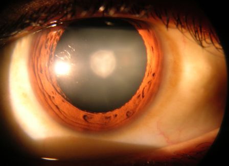
Cataract in Human Eye. Magnified view seen on examination with a slit lamp
-
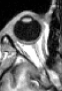
MRI scan of human eye showing lens.
-
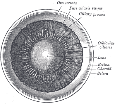
Interior of anterior chamber of eye.
-
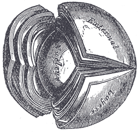
The crystalline lens, hardened and divided.
-
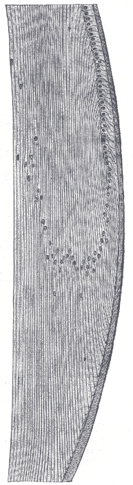
Section through the margin of the lens, showing the transition of the epithelium into the lens fibers.


References
- ↑ John Forrester, Andrew Dick, Paul McMenamin, William Lee (1996). The Eye: Basic Sciences in Practice. London: W.B. Saunders Company Ltd. p. 28 ISBN 0-7020-1790-6
- ↑ The Eye: Basic Sciences in Practice, p. 28, ISBN 0-7020-1790-6
- ↑ The Eye: Basic Sciences in Practice, p. 28, ISBN 0-7020-1790-6
- ↑ The Eye: Basic Sciences in Practice, p. 28, ISBN 0-7020-1790-6
- ↑ O. Candia (2004). Electrolyte and fluid transport across corneal, conjunctival and lens epithelia. Experimental Eye Research 78 (3): 527-535.
- ↑ The Eye: Basic Sciences in Practice, p. 28, ISBN 0-7020-1790-6
- ↑ Hecht, Eugene. Optics, 2nd ed. (1987), Addison Wesley, ISBN 0-201-11609-X. p. 178.
- ↑ W. Hoehenwarter, J. Klose and P. R. Jungblut (2006). Eye lens proteomics. Amino Acids 30(4): 369-389.
- ↑ U. Andley (2006). Crystallins in the eye: function and pathology. Progress in Retinal and Eye Research 26 (1): 78-98.
- ↑ U. Andley (2006). Crystallins in the eye: function and pathology. Progress in Retinal and Eye Research 26 (1): 78-98.
- ↑ H Bloemendal, W. de Jong, R Jaenicke, NH Lubsen, C Slingsby and A Tardieu (2004). Aging and vision: structure, stability, and function of lens crystallins. Progress in Biophysics and Molecular Biology 86 (3): 407-485.
- ↑ The Eye: Basic Sciences in Practice, p. 102, ISBN 0-7020-1790-6
- ↑ The Eye: Basic Sciences in Practice, p. 102, ISBN 0-7020-1790-6
- ↑ U. Andley (2006). Crystallins in the eye: function and pathology. Progress in Retinal and Eye Research 26 (1): 78-98.
- ↑ The Eye: Basic Sciences in Practice, p. 28, ISBN 0-7020-1790-6
- ↑ Whikehart, David R. (2003). Biochemistry of the Eye, 2nd ed. 2003. Philadelphia: Butterworth Heinemann, p.107-8 ISBN 0-7506-7152-1
- ↑ The Eye: Basic Sciences in Practice, p. 102, ISBN 0-7020-1790-6
- ↑ The Eye: Basic Sciences in Practice, p. 104, ISBN 0-7020-1790-6
- ↑ O. Candia (2004). Electrolyte and fluid transport across corneal, conjunctival and lens epithelia.Experimental Eye Research 78 (3): 527-535.
- ↑ Biochemistry of the Eye, 2nd ed, p.107-8, ISBN 0-7506-7152-1
- ↑ Biochemistry of the Eye, 2nd ed, p.107-8, ISBN 0-7506-7152-1
- ↑ Biochemistry of the Eye, 2nd ed, p.107-8, ISBN 0-7506-7152-1
See also
External links
- Histology image: 08001loa – Histology Learning System at Boston University
bn:লেন্স (শারীরবিদ্যা) ca:Cristal·lí cs:Čočka (oko) de:Linse (Auge) et:Silmalääts eo:Kristalino eu:Kristalino gl:Cristalino it:Cristallino lt:Lęšiukas nl:Ooglens sk:Šošovka (anatómia) fi:Mykiö uk:Кришталик yi:לענס