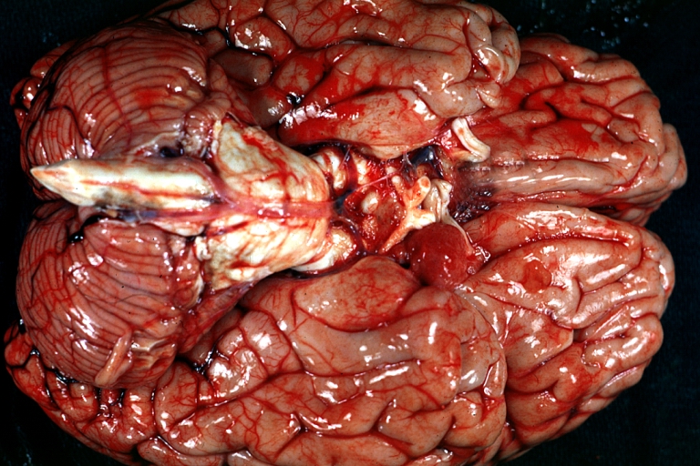
Intracranial berry aneurysm
| Intracranial berry aneurysm | |
 | |
|---|---|
| Brain: Berry Aneurysm: Gross base of brain with large unruptured berry aneurysm. Image courtesy of Professor Peter Anderson DVM PhD and published with permission © PEIR, University of Alabama at Birmingham, Department of Pathology |
Editor-In-Chief: C. Michael Gibson, M.S., M.D. [1]
Synonyms and keywords: Berry aneursym; saccular aneurysm
Overview
An intracranial berry aneurysm, also known as a saccular aneurysm, is a sac-like outpouching in a cerebral blood vessel, which can seem berry-shaped, hence the name. Once a berry aneurysm has formed it is likely to rupture, causing a stroke. Thus they are serious medical emergencies, and should be treated as soon as possible.
Berry aneurysms are due to a combination of hypertension and weakness of the blood vessel wall. Weak or thinned parts of the cerebral vasculature are vulnerable to the increased hydrostatic pressure caused by hypertension, and will bulge out. Some parts of the brain vasculature are inherently weak: particularly areas along the Circle of Willis, where small communicating vessels link the main cerebral vessels. These areas are particularly susceptible to berry aneurysms.[1] These aneurysms can form in both the anterior circulation and the posterior circulation in the brain. Typical sites include the junctures or bifurcation of the major cerebral blood vessels: the internal (anterior circulation), and the vertebral artery and basilar artery (posterior circulation).
Intracranial berry aneurysms are the most common kind of aneurysm in the brain. Their incidence is 1 in 1000 people per year (around 27,000 cases per year in the United States). They have a mortality rate of 70-90%.
The typical symptom of a ruptured berry aneurysm is the development of a headache so bad that the patient says "it is the worst headache I have ever had."[2]
The risk factors for developing berry aneurysms include any condition that causes hypertension, or any condition that causes weakening of blood vessel walls.[3] Conditions causing hypertension include atherosclerosis, renal disease, vasculitis, or drugs that induce hypertension (such as cocaine). Conditions that cause weakening of blood vessel walls include gentical or acquired malformation of a blood vessel, genetic disorders of connective tissue, head trauma, and infections.
Complications of a ruptured aneurysm include stroke, vasospasm, and subarachnoid hemorrhage. Subarachnoid hemorrhage (bleeding in the space between the bones of the skull and the brain) can lead to hydrocephalus.
Signs and Symptoms
Most intracranial berry aneurysms do not show any symptoms unless they have ruptured, which causes bleeding in the brain. Small aneurysms that maintain their size generally will not show symptoms. But larger aneurysms that are steadily growing can put pressure on nerves and tissues. Some signs of an unruptured aneurysm are:
- Headaches
- Double vision (Diplopia)
- Loss of vision
- Eye and neck pain
- Sentinel or warning headaches. These are headaches that are caused by leakage of blood into the brain for days up to weeks prior to the aneurysm's rupture (only a small percentage of patients experience a sentinel headache before rupture).
Symptoms of a ruptured aneurysm are:
- Sudden, severe headache
- Lethargy
- Confusion and Stupor
- Seizures
- Sudden mood swings (Impulsivity, Irritability, Poor temper control)
- Speech impediment (dysarthria)
- Eyelids dropping (Ptosis)
Diagnosis and Treatment
Once suspected, berry aneurysms can be diagnosed using angiography, Magnetic Resonance Imaging, CT scans, and Cerebrospinal fluid analysis.
Most cerebral aneurysms are unobserved until they have already ruptured. Diagnostic tests can be used to detect if an aneurysm has or will rupture. Tests are usually acquired after a subarachnoid hemorrhage, to confirm the presence of an aneurysm.
If an unruptured aneurysm has been discovered in the brain of a patient, there are surgical procedures that can treat the aneurysm. One of them is Micro-Vascular Clipping, where the surgeon goes into the brain and cuts off blood flow to the aneurysm. Once this surgery is performed, the clip remains in the patient and prevents any future bleeding. It has been proven to be highly effective, because most aneurysms that are clipped do not return.
Another, related procedure is called Occlusion. In this surgical procedure, the entire artery involved with an aneurysm is clamped off (occluded). After this is done, a small blood vessel is used to reroute the blood away from the afflicted artery. There are also other forms of treatment for berry aneurysms.
People who have been diagnosed with a berry aneurysm, or any kind of aneurysm, should take steps to control high blood pressure, including smoking cessation and avoidance of cocaine and other drugs that elevate blood pressure.