Foramen ovale (heart)

You don't need to be Editor-In-Chief to add or edit content to WikiDoc. You can begin to add to or edit text on this WikiDoc page by clicking on the edit button at the top of this page. Next enter or edit the information that you would like to appear here. Once you are done editing, scroll down and click the Save page button at the bottom of the page.

Jump to: navigation, search
Foramen ovale (heart)
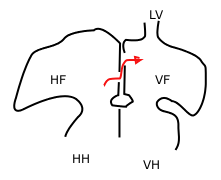
Sketch showing foramen ovale in a fetal heart. Red arrow shows blood from the inferior caval vein. HV: right atrium, VF: left atrium. HH og VH: right and left ventricle. The heart still has a common pulmonary vein (LV), in stead of four.
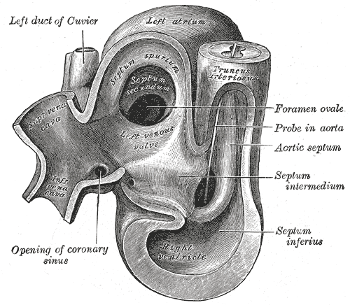
Heart of human embryo of about thirty-five days, opened on right side.
Gray's subject #135 512
MeSH Foramen+Ovale

WikiDoc Resources for

Foramen ovale (heart)

Articles

Most recent articles on Foramen ovale (heart)

Most cited articles on Foramen ovale (heart)

Review articles on Foramen ovale (heart)

Articles on Foramen ovale (heart) in N Eng J Med, Lancet, BMJ

Media

Powerpoint slides on Foramen ovale (heart)

Images of Foramen ovale (heart)

Photos of Foramen ovale (heart)

Podcasts & MP3s on Foramen ovale (heart)

Videos on Foramen ovale (heart)

Evidence Based Medicine

Cochrane Collaboration on Foramen ovale (heart)

Bandolier on Foramen ovale (heart)

TRIP on Foramen ovale (heart)

Clinical Trials

Ongoing Trials on Foramen ovale (heart) at Clinical Trials.gov

Trial results on Foramen ovale (heart)

Clinical Trials on Foramen ovale (heart) at Google

Guidelines / Policies / Govt

US National Guidelines Clearinghouse on Foramen ovale (heart)

NICE Guidance on Foramen ovale (heart)

NHS PRODIGY Guidance

FDA on Foramen ovale (heart)

CDC on Foramen ovale (heart)

Books

Books on Foramen ovale (heart)

News

Foramen ovale (heart) in the news

Be alerted to news on Foramen ovale (heart)

News trends on Foramen ovale (heart)

Commentary

Blogs on Foramen ovale (heart)

Definitions

Definitions of Foramen ovale (heart)

Patient Resources / Community

Patient resources on Foramen ovale (heart)

Discussion groups on Foramen ovale (heart)

Patient Handouts on Foramen ovale (heart)

Directions to Hospitals Treating Foramen ovale (heart)

Risk calculators and risk factors for Foramen ovale (heart)

Healthcare Provider Resources

Symptoms of Foramen ovale (heart)

Causes & Risk Factors for Foramen ovale (heart)

Diagnostic studies for Foramen ovale (heart)

Treatment of Foramen ovale (heart)

Continuing Medical Education (CME)

CME Programs on Foramen ovale (heart)

International

Foramen ovale (heart) en Espanol

Foramen ovale (heart) en Francais

Business

Foramen ovale (heart) in the Marketplace

Patents on Foramen ovale (heart)

Experimental / Informatics

List of terms related to Foramen ovale (heart)

Editor-In-Chief: C. Michael Gibson, M.S., M.D. [1] Phone:617-632-7753

Please Take Over This Page and Apply to be Editor-In-Chief for this topic: There can be one or more than one Editor-In-Chief. You may also apply to be an Associate Editor-In-Chief of one of the subtopics below. Please mail us [2] to indicate your interest in serving either as an Editor-In-Chief of the entire topic or as an Associate Editor-In-Chief for a subtopic. Please be sure to attach your CV and or biographical sketch.

Overview

In the fetal heart, the foramen ovale (or ostium secundum of Born) allows blood to enter the left atrium from the right atrium. It is one of two shunts, the other being the ductus arteriosus, that allows blood entering the right atrium to bypass the pulmonary circulation. Another similar adaptation in the fetus is the ductus venosus. In most individuals, the foramen ovale closes within the first year after birth to form the fossa ovalis.

Development

The foramen ovale begins forming late in the fourth week of gestation. Initially the atria are separated from one another by the septum primum except for a small opening in the septum, the ostium primum. As the septum primum grows, the ostium primum narrows and eventually closes. Before it does so, bloodflow from the inferior vena cava wears down a portion of the septum primum, forming the ostium secundum. Some embryologists postulate that the ostium secundum may be formed through programmed cell death. The ostium secundum provides communication between the atria after the ostium primum closes completely. Subsequently, a second wall of tissue, the septum secundum, grows over the ostium secundum in the right atrium. Bloodflow then only passes from the right to left atrium by way of a small passageway in the septum secundum and then through the ostium secundum. This passageway is called the foramen ovale.

Closure

Normally this opening closes in the first year of life. When the lungs become functional at birth, the pulmonary pressure decreases and the left atrial pressure exceeds that of the right. This forces the septum primum against the septum secundum, functionally closing the foramen ovale. In time the septa eventually fuse, leaving a remnant of the foramen ovale, the fossa ovalis.

Clinical relevance

In about 30% of adults the foramen ovale does not close completely, but remains as a small patent foramen ovale. People with a patent foramen ovale are more likely to suffer from migraine headaches. Corrective surgery often results in the complete cessation of migraines in some patients, and a greater than 50% reduction in migraines on average [3].

References

  • Carlson, Bruce (2004). Human Embryology And Developmental Biology (3rd edition). Elsevier Mosby. ISBN 0-323-03649-X. 

de:Foramen ovale nl:Foramen ovale nn:Foramen ovalesv:Foramen ovale


WikiDoc Help Menu

Quick Start..

Editing basics

Advanced editing

Communicating your edits

Help Videos You Can Watch

Acknowledgement and Attribution Regarding Sources of Content

Some of the initial content on this page may be incorporated in part from copyleft sources in the public domain including wikis such as Wikipedia and AskDrWiki. Drug information for patients came from the The National Library of Medicine. Infectious disease information may have come from the Centers for Disease Control (CDC). Differential Diagnoses are drawn from clinicians as well as an amalgamation of 3 sources: 1.The Disease Database; 2. Kahan, Scott, Smith, Ellen G. In A Page: Signs and Symptoms. Malden, Massachusetts: Blackwell Publishing, 2004:3; 3. Sailer, Christian, Wasner, Susanne. Differential Diagnosis Pocket. Hermosa Beach, CA: Borm Bruckmeir Publishing LLC, 2002:7 .

In other languages