Cerebral hypoxia other imaging findings
Jump to navigation
Jump to search
|
Cerebral hypoxia Microchapters |
|
Diagnosis |
|---|
|
Treatment |
|
Case Studies |
|
Cerebral hypoxia other imaging findings On the Web |
|
American Roentgen Ray Society Images of Cerebral hypoxia other imaging findings |
|
Risk calculators and risk factors for Cerebral hypoxia other imaging findings |
Editor-In-Chief: C. Michael Gibson, M.S., M.D. [1]
Please help WikiDoc by adding content here. It's easy! Click here to learn about editing.
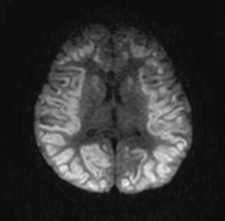
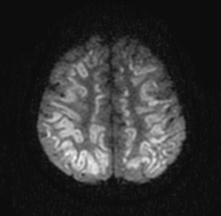
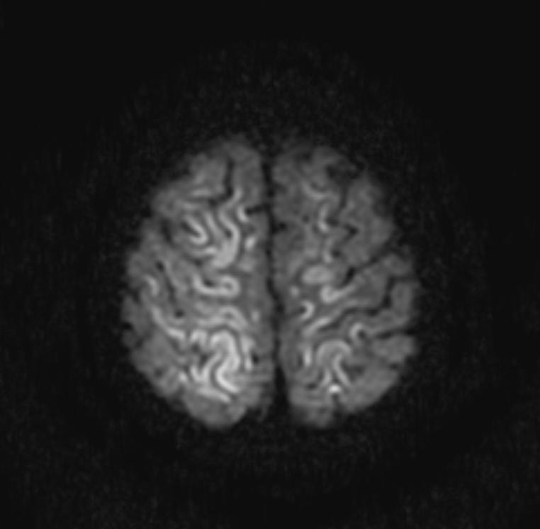
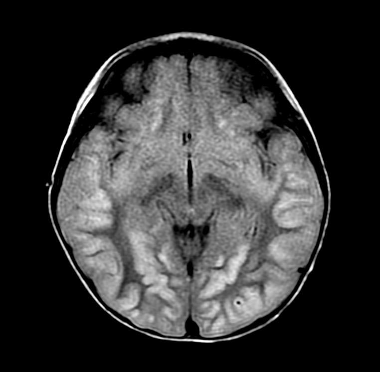
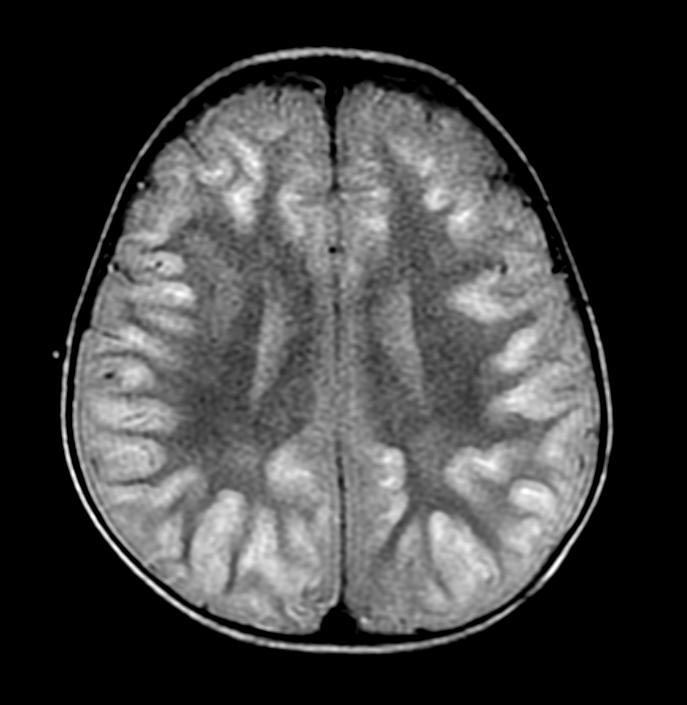
Diagnostic Findings
- Diffuse loss of gray/white differentiation
- Diffuse sulcal effacement
Patient #1:


Patient #2:
-
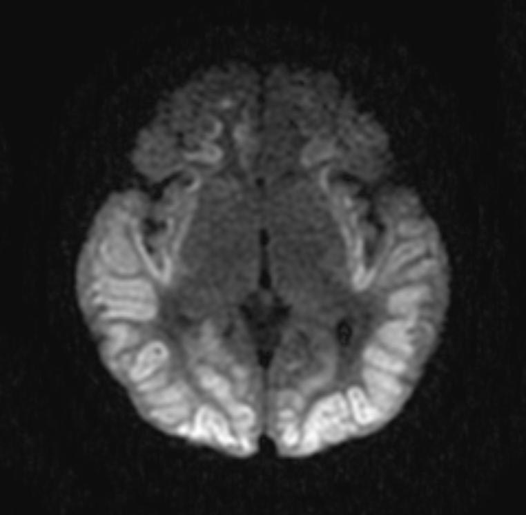 DWI
DWI -
 DWI
DWI


-
 DWI
DWI -
 DWI
DWI


-
 FLAIR
FLAIR -
 FLAIR
FLAIR -
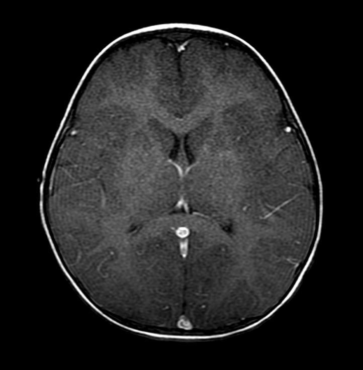 T1 with GAD


