Anatomy of the heart right ventricle
Jump to navigation
Jump to search
|
Anatomy of the heart Microchapters |
|
Anatomy of the heart right ventricle On the Web |
|---|
|
American Roentgen Ray Society Images of Anatomy of the heart right ventricle |
|
Risk calculators and risk factors for Anatomy of the heart right ventricle |
Editor-In-Chief: C. Michael Gibson, M.S., M.D. [1]; Assistant Editor(s)-in-Chief: Rim Halaby, Yazan Daaboul
Overview
Right ventricle is a triangular chamber extending from the right atrium to the cardiac apex and receives deoxygenated blood from the right atrium.
Right Ventricle
- The right ventricle forms almost the entire inferior border of the heart, the largest part of the anterior surface of the heart, and also contributes to the diaphragmatic surface.
- The interior of the right ventricle has irregular muscle elevations called trabeculae carneae.
- The flow of blood in the right ventricle:
- The inflow of blood into the right ventricle enters first posteriorly.
- The outflow of blood leaves superiorly and to the left at the level of the pulmonary trunk.
- Therefore, blood takes a U-shaped path through the right ventricle in a total tract length of 2 cm.
Tricuspid Valve
- The inflow part receives deoxygenated blood from the right atrium through the right atrioventricular or tricuspid, orifice via the tricuspid valve.
- The orifice is a located retro-sternally between fourth and fifth intercostal spaces.
- It is characterized by a unique fibrous ring that resists dilation due to pressure variations.
- The tricuspid valve contains 3 cusps: anterior, posterior, and septal.
- The valve cusps play an important role in preventing blood regurgitation from the right ventricle to the right atrium during ventricular contraction.
- Hence when blood is forced through the right atrio-ventricular orifice, the tricuspid valve cusps are pushed aside.
- Below is an image Courtesy of Professor Peter Anderson DVM PhD and Published with permission. © PEIR, University of Alabama at Birmingham, Department of Pathology
-
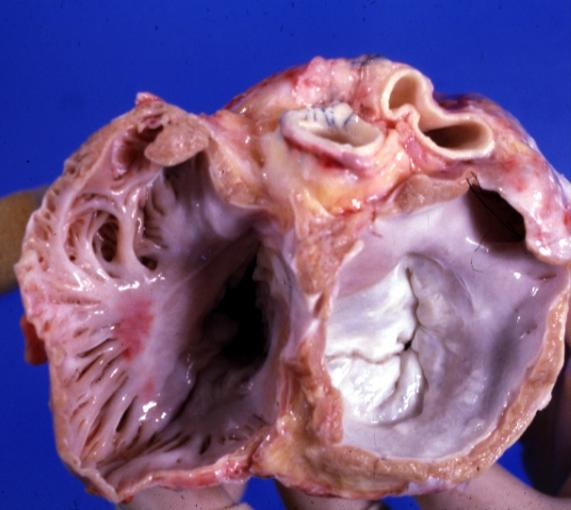 Mitral and tricuspid valves from atria. Normal valvular anatomy

Tendinous Chords
- All tricuspid valve cusps are attached on their free edges and ventricular surfaces via delicate tendons called tendinous chords (L. chordae tendineae) on their free edges; with each chord attaching simultaneously 2 adjacent valve cusps.
- The tendinous chords function in preventing valvular prolapse when ventricular pressure rises due to ventricular contraction.
Papillary Muscles
- Papillary muscles are three projections whose apices give rise to tendinous chords.
- Their bases are attached to the wall of the right ventricle.
- With respect to the right ventricle, they are located anteriorly, posteriorly, and septally and hence named accordingly.
- The anterior papillary muscle is the largest, while the septal is the smallest and mayb be multiple.
- Significantly, papillary muscles contract before contraction of the right ventricular in order to tighten the tendinous chords. As such, the tricuspid valve closes by drawing the cusps together before ventricular contraction and blood is prevented from regurgitation to the right atrium.
Interventricular Septum
- Interventricular septum is a strong oblique wall between the right and left ventricles. It is composed of two parts:
- Membranous: thin, superoposterior part and continuous with the fibrous skeleton of the heart.
- Muscular: thick bulge into the cavity of the right ventricle. The prominent bulging is due to the overwhelming pressure in the left ventricle with respect to its counterpart in the right side.
Septomarginal Trabecula (Moderator Band)
- The septomarginal trabecula is a curved muscular bundle running from the inferior part of the inter-ventricular septum to the base of the anterior papillary muscle.
- It carries part of the right bundle branches of the AV bundle, necessary for electrical conduction, to the anterior papillary muscle of the right ventricle.
- It is believed that via this conduction route that the anterior papillary muscle achieves its coordinated contraction.
Pulmonary Valve
- The pulmonary valve is located at the apex of the conus arteriosus (infundibulum), a smooth-walled, cone-shaped portion of the right ventricle inferior to the opening of the pulmonary trunk (pulmonary artery).
- It is at the level of the left third costal cartilage.
- The pulmonary valve contains three semilunar cusps: anterior, right, and left.
- Each semilunar valve is concave when viewed superiorly and has three structures: one fibrous nodule and two lunules.
- The nodule and the lunules help sealing the valve cusps a prevent backflow of blood during diastole.
Pulmonary Sinus and Pulmonary Trunk
- Pulmonary sinuses are the spaces at the origin of the pulmonary trunk between the dilated wall of the vessel and each cusp of the pulmonary valve.
- Blood in the pulmonary sinuses prevents sticking of the cusps to the walls of the pulmonary trunk and consequential failure to close.