Adrenal hemorrhage echocardiography or ultrasound
Jump to navigation
Jump to search
|
Adrenal hemorrhage Microchapters |
|
Diagnosis |
|---|
|
Treatment |
|
Case Studies |
|
Adrenal hemorrhage echocardiography or ultrasound On the Web |
|
American Roentgen Ray Society Images of Adrenal hemorrhage echocardiography or ultrasound |
|
Adrenal hemorrhage echocardiography or ultrasound in the news |
|
Risk calculators and risk factors for Adrenal hemorrhage echocardiography or ultrasound |
Editor-In-Chief: C. Michael Gibson, M.S., M.D. [1]
Overview
Ultrasonography
- Pattern of echogenicity of an adrenal hematoma depends on its age
- Early-stage hematoma appears solid with diffuse or inhomogeneous echogenicity.
- As liquefaction occurs, the mass demonstrates mixed echogenicity with a central hypoechoic region and eventually becomes completely anechoic and cystlike.
- Calcifications may be seen in the walls of the hematoma as early as 1–2 weeks after onset and gradually compact as the blood is absorbed.
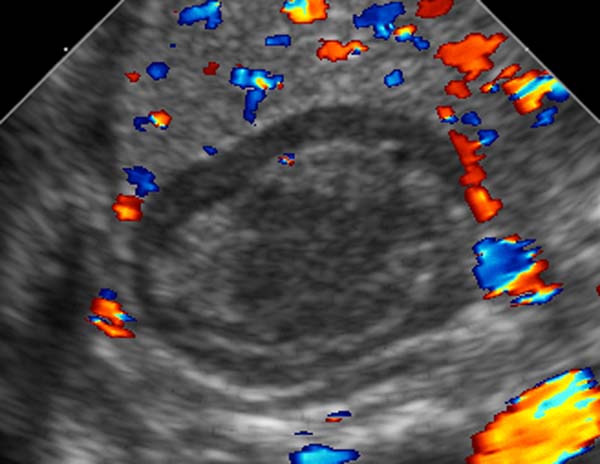
- Color Doppler and power Doppler imaging allow confirmation of the avascular nature of the mass.
-
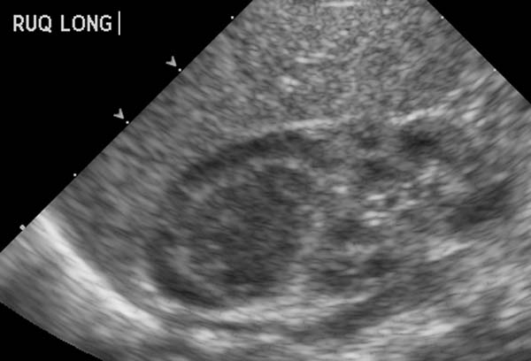 Ultrasonography: Adrenal hemorrhage
 -
 Ultrasonography: Adrenal hemorrhage

