Warthin's tumor biopsy: Difference between revisions
No edit summary |
Nazia Fuad (talk | contribs) No edit summary |
||
| (9 intermediate revisions by 2 users not shown) | |||
| Line 1: | Line 1: | ||
__NOTOC__ | __NOTOC__ | ||
{{Warthin's tumor}} | {{Warthin's tumor}} | ||
{{CMG}} | {{CMG}}; {{AE}} {{Ammu}} | ||
==Overview== | ==Overview== | ||
== | On [[biopsy]], Warthin's tumor is characterized by [[cystic]] spaces surrounded by two uniform rows of [[cell (biology)|cells]] with centrally placed pyknotic [[cell nucleus|nuclei]], [[papillae]] with a two rows of pink [[epithelial cells]], and [[lymphoid]] [[stroma]]. | ||
Salivary gland [[biopsy]] | ==Biopsy== | ||
[[Salivary gland]] [[biopsy]] is the most important test for diagnosis of Warthin's tumor. | |||
==Microscopic Pathology== | |||
* The appearance of this tumor under the microscope is unique. There are [[cystic]] spaces surrounded by two uniform rows of [[cell (biology)|cells]] with centrally placed pyknotic [[cell nucleus|nuclei]]. | |||
* The [[cystic]] spaces have [[epithelium]] referred to as [[papillary]] infoldings that protude into them. Additionally, the [[epithelium]] has [[Stroma|lymphoid stroma]] with [[germinal center]] formation. | |||
* [[Papillae]] (nipple-shaped structures) with a two rows of pink ([[eosinophilic]]) [[epithelial cells]] (with cuboidal [[Basal cell|basal cells]] and columnar luminal cells) | |||
* Fibrous capsule - pink & [[homogenous]] on H&E stain | |||
* Cystic space filled with debris in situ (not [[necrosis]]) | |||
* [[Lymphoid]] [[stroma]] | |||
* Additionally, the [[epithelium]] has lymphoid [[stroma]] with [[germinal center]] formation.<ref>Warthin's tumor librepathology (2015) http://librepathology.org/wiki/index.php/Warthin_tumour Accessed on December 14, 2015</ref> | |||
<gallery> | |||
Image:Warthin tumor (1).jpg|Histopathology of Warthin tumor in the parotid gland. H&E stain<ref name="AbidStack2014">{{cite journal|last1=Abid|first1=Syed A.|last2=Stack|first2=Brendan C.|last3=Bodenner|first3=Donald L.|title=Metastatic Follicular Thyroid Carcinoma Secreting Thyroid Hormone and Radioiodine Avid without Stimulation: A Case Report and Literature Review|journal=Case Reports in Endocrinology|volume=2014|year=2014|pages=1–6|issn=2090-6501|doi=10.1155/2014/584513}}</ref> | |||
Image:Warthin tumor (2).jpg|Histopathology of Warthin tumor in the parotid gland. Another view of a file "Warthin tumor (1).jpg". H&E stain..<ref name="AbidStack2014">{{cite journal|last1=Abid|first1=Syed A.|last2=Stack|first2=Brendan C.|last3=Bodenner|first3=Donald L.|title=Metastatic Follicular Thyroid Carcinoma Secreting Thyroid Hormone and Radioiodine Avid without Stimulation: A Case Report and Literature Review|journal=Case Reports in Endocrinology|volume=2014|year=2014|pages=1–6|issn=2090-6501|doi=10.1155/2014/584513}}</ref> | |||
Image:Warthin tumor (3).jpg|Histopathology of Warthin tumor in the parotid gland. Higher magnification of a file "Warthin tumor (1).jpg". H&E stain.<ref name="AbidStack2014">{{cite journal|last1=Abid|first1=Syed A.|last2=Stack|first2=Brendan C.|last3=Bodenner|first3=Donald L.|title=Metastatic Follicular Thyroid Carcinoma Secreting Thyroid Hormone and Radioiodine Avid without Stimulation: A Case Report and Literature Review|journal=Case Reports in Endocrinology|volume=2014|year=2014|pages=1–6|issn=2090-6501|doi=10.1155/2014/584513}}</ref> | |||
Image:Papillary cystadenoma lymphomato 01.jpg|Histopathology of Warthin tumor in the parotid gland. Image courtesy of Nephron [http://www.librepathology.org librepathology] (original file [http://librepathology.org/wiki/index.php/File:Papillary_cystadenoma_lymphomatosum1.jpg ‘’here’’]). [http://librepathology.org/licence Creative Commons BYSANC] | |||
Image:Papillary cystadenoma lymphomato 02.jpg|Histopathology of Warthin tumor in the parotid gland. Image courtesy of Nephron [http://www.librepathology.org librepathology] (original file [http://librepathology.org/wiki/index.php/File:Papillary_cystadenoma_lymphomatosum2.jpg ‘’here’’]). [http://librepathology.org/licence Creative Commons BYSANC] | |||
Image:Papillary cystadenoma lymphomato 02.jpg|Histopathology of Warthin tumor in the parotid gland. Image courtesy of Nephron [http://www.librepathology.org librepathology] (original file [http://librepathology.org/wiki/index.php/File:Papillary_cystadenoma_lymphomatosum2.jpg ‘’here’’]). [http://librepathology.org/licence Creative Commons BYSANC] | |||
Image:777px-Papillary cystadenoma lymphomato 03.jpg|Histopathology of Warthin tumor in the parotid gland. Image courtesy of Nephron [http://www.librepathology.org librepathology] (original file [http://librepathology.org/wiki/index.php/File:Papillary_cystadenoma_lymphomatosum2.jpg ‘’here’’]). [http://librepathology.org/licence Creative Commons BYSANC] | |||
</gallery> | |||
==References== | ==References== | ||
| Line 14: | Line 32: | ||
[[Category:Oral pathology]] | [[Category:Oral pathology]] | ||
[[Category:Types of cancer]] | [[Category:Types of cancer]] | ||
[[Category:Disease]] | [[Category:Disease]] | ||
[[Category:Up-To-Date]] | |||
[[Category:Oncology]] | |||
[[Category:Medicine]] | |||
[[Category:Otolaryngology]] | |||
[[Category:Gastroenterology]] | |||
Latest revision as of 18:16, 5 December 2018
|
Warthin's tumor Microchapters |
|
Diagnosis |
|---|
|
Treatment |
|
Case Studies |
|
Warthin's tumor biopsy On the Web |
|
American Roentgen Ray Society Images of Warthin's tumor biopsy |
|
Risk calculators and risk factors for Warthin's tumor biopsy |
Editor-In-Chief: C. Michael Gibson, M.S., M.D. [1]; Associate Editor(s)-in-Chief: Ammu Susheela, M.D. [2]
Overview
On biopsy, Warthin's tumor is characterized by cystic spaces surrounded by two uniform rows of cells with centrally placed pyknotic nuclei, papillae with a two rows of pink epithelial cells, and lymphoid stroma.
Biopsy
Salivary gland biopsy is the most important test for diagnosis of Warthin's tumor.
Microscopic Pathology
- The appearance of this tumor under the microscope is unique. There are cystic spaces surrounded by two uniform rows of cells with centrally placed pyknotic nuclei.
- The cystic spaces have epithelium referred to as papillary infoldings that protude into them. Additionally, the epithelium has lymphoid stroma with germinal center formation.
- Papillae (nipple-shaped structures) with a two rows of pink (eosinophilic) epithelial cells (with cuboidal basal cells and columnar luminal cells)
- Fibrous capsule - pink & homogenous on H&E stain
- Cystic space filled with debris in situ (not necrosis)
- Lymphoid stroma
- Additionally, the epithelium has lymphoid stroma with germinal center formation.[1]
-
![Histopathology of Warthin tumor in the parotid gland. H&E stain[2]](/images/c/c7/Warthin_tumor_%281%29.jpg)
Histopathology of Warthin tumor in the parotid gland. H&E stain[2]
-
![Histopathology of Warthin tumor in the parotid gland. Another view of a file "Warthin tumor (1).jpg". H&E stain..[2]](/images/c/cb/Warthin_tumor_%282%29.jpg)
Histopathology of Warthin tumor in the parotid gland. Another view of a file "Warthin tumor (1).jpg". H&E stain..[2]
-
![Histopathology of Warthin tumor in the parotid gland. Higher magnification of a file "Warthin tumor (1).jpg". H&E stain.[2]](/images/d/d2/Warthin_tumor_%283%29.jpg)
Histopathology of Warthin tumor in the parotid gland. Higher magnification of a file "Warthin tumor (1).jpg". H&E stain.[2]
-
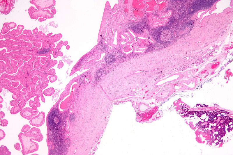
Histopathology of Warthin tumor in the parotid gland. Image courtesy of Nephron librepathology (original file ‘’here’’). Creative Commons BYSANC
-
Histopathology of Warthin tumor in the parotid gland. Image courtesy of Nephron librepathology (original file ‘’here’’). Creative Commons BYSANC
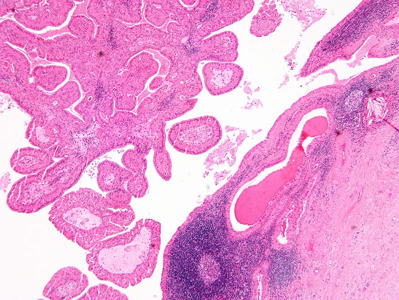
-
Histopathology of Warthin tumor in the parotid gland. Image courtesy of Nephron librepathology (original file ‘’here’’). Creative Commons BYSANC
-
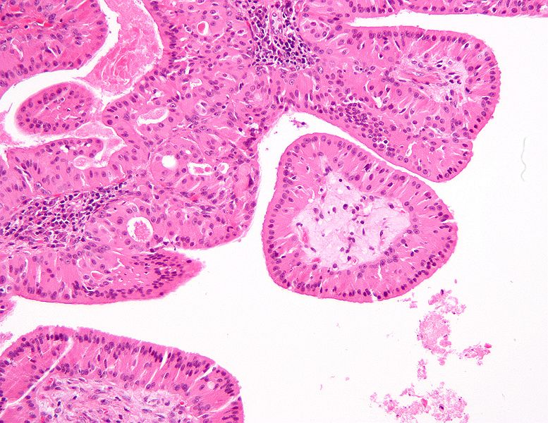
Histopathology of Warthin tumor in the parotid gland. Image courtesy of Nephron librepathology (original file ‘’here’’). Creative Commons BYSANC
![Histopathology of Warthin tumor in the parotid gland. H&E stain[2]](/index.php/File:Warthin_tumor_(1).jpg)
![Histopathology of Warthin tumor in the parotid gland. Another view of a file "Warthin tumor (1).jpg". H&E stain..[2]](/index.php/File:Warthin_tumor_(2).jpg)
![Histopathology of Warthin tumor in the parotid gland. Higher magnification of a file "Warthin tumor (1).jpg". H&E stain.[2]](/index.php/File:Warthin_tumor_(3).jpg)


References
- ↑ Warthin's tumor librepathology (2015) http://librepathology.org/wiki/index.php/Warthin_tumour Accessed on December 14, 2015
- ↑ 2.0 2.1 2.2 Abid, Syed A.; Stack, Brendan C.; Bodenner, Donald L. (2014). "Metastatic Follicular Thyroid Carcinoma Secreting Thyroid Hormone and Radioiodine Avid without Stimulation: A Case Report and Literature Review". Case Reports in Endocrinology. 2014: 1–6. doi:10.1155/2014/584513. ISSN 2090-6501.