Waldenström's macroglobulinemia bone marrow aspiration and biopsy: Difference between revisions
Sara Mohsin (talk | contribs) |
No edit summary |
||
| Line 11: | Line 11: | ||
Findings suggestive of Waldenström macroglobulinemia include:<ref name="pmid18555588">{{cite journal| author=Leleu X, Roccaro AM, Moreau AS, Dupire S, Robu D, Gay J et al.| title=Waldenstrom macroglobulinemia. | journal=Cancer Lett | year= 2008 | volume= 270 | issue= 1 | pages= 95-107 | pmid=18555588 | doi=10.1016/j.canlet.2008.04.040 | pmc=3133633 | url=https://www.ncbi.nlm.nih.gov/entrez/eutils/elink.fcgi?dbfrom=pubmed&tool=sumsearch.org/cite&retmode=ref&cmd=prlinks&id=18555588 }} </ref> | Findings suggestive of Waldenström macroglobulinemia include:<ref name="pmid18555588">{{cite journal| author=Leleu X, Roccaro AM, Moreau AS, Dupire S, Robu D, Gay J et al.| title=Waldenstrom macroglobulinemia. | journal=Cancer Lett | year= 2008 | volume= 270 | issue= 1 | pages= 95-107 | pmid=18555588 | doi=10.1016/j.canlet.2008.04.040 | pmc=3133633 | url=https://www.ncbi.nlm.nih.gov/entrez/eutils/elink.fcgi?dbfrom=pubmed&tool=sumsearch.org/cite&retmode=ref&cmd=prlinks&id=18555588 }} </ref> | ||
* A hypercellular bone marrow aspirate. | * A hypercellular bone marrow aspirate. | ||
* Lymphoplasmacytic infiltrate with characteristic immunophenotype. | * Lymphoplasmacytic infiltrate with characteristic [[immunophenotype]]. | ||
{| | {| | ||
| Line 24: | Line 24: | ||
* Hypercellular and [[Infiltration (medical)|infiltrated]] with [[lymphoid]] and [[Plasmacytoid|plasmacytoid cells]]. | * Hypercellular and [[Infiltration (medical)|infiltrated]] with [[lymphoid]] and [[Plasmacytoid|plasmacytoid cells]]. | ||
* Dutcher bodies (PAS positive intra-nuclear vacuoles containing IgM monoclonal protein). | * Dutcher bodies (PAS positive intra-nuclear vacuoles containing [[IgM]] monoclonal protein). | ||
** Characteristic feature of Waldenström macroglobulinemia. | ** Characteristic feature of Waldenström macroglobulinemia. | ||
Three patterns of marrow involvement are described, as follows: | Three patterns of marrow involvement are described, as follows: | ||
* Lymphoplasmacytoid cells (lymphoplasmacytic and small lymphocytes) in a nodular pattern. | * Lymphoplasmacytoid cells ([[Lymphoplasmacytic lymphoma|lymphoplasmacytic]] and small lymphocytes) in a nodular pattern. | ||
* Lymphoplasmacytic cells (small lymphocytes, mature plasma cells, mast cells) in an interstitial/nodular pattern. | * Lymphoplasmacytic cells (small lymphocytes, mature plasma cells, mast cells) in an interstitial/nodular pattern. | ||
* A polymorphous infiltrate (small lymphocytes, plasma cells, plasmacytoid cells, immunoblasts with mitotic figures). | * A polymorphous infiltrate (small lymphocytes, plasma cells, plasmacytoid cells, immunoblasts with mitotic figures). | ||
Revision as of 18:31, 8 April 2019
|
Waldenström's macroglobulinemia Microchapters |
|
Differentiating Waldenström's macroglobulinemia from other Diseases |
|---|
|
Diagnosis |
|
Treatment |
|
Case Studies |
|
Waldenström's macroglobulinemia bone marrow aspiration and biopsy On the Web |
|
American Roentgen Ray Society Images of Waldenström's macroglobulinemia bone marrow aspiration and biopsy |
|
FDA on Waldenström's macroglobulinemia bone marrow aspiration and biopsy |
|
CDC on Waldenström's macroglobulinemia bone marrow aspiration and biopsy |
|
Waldenström's macroglobulinemia bone marrow aspiration and biopsy in the news |
|
Blogs on Waldenström's macroglobulinemia bone marrow aspiration and biopsy |
|
Directions to Hospitals Treating Waldenström's macroglobulinemia |
Editor-In-Chief: C. Michael Gibson, M.S., M.D. [1]; Associate Editor(s)-in-Chief: Sara Mohsin, M.D.[2] Roukoz A. Karam, M.D.[3]
Overview
A bone marrow aspiration is essential in the diagnosis of Waldenström macroglobulinemia.
Bone Marrow Aspirate
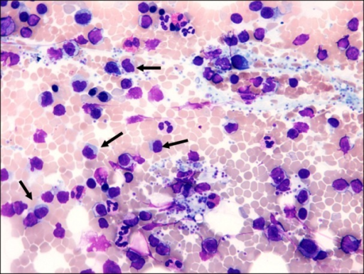
A bone marrow aspirate is essential in the diagnosis of Waldenström macroglobulinemia.
Findings suggestive of Waldenström macroglobulinemia include:[1]
- A hypercellular bone marrow aspirate.
- Lymphoplasmacytic infiltrate with characteristic immunophenotype.
 |
Bone Marrow Biopsy
A bone marrow biopsy may be helpful in the diagnosis of Waldenström macroglobulinemia. [1]
Findings on the biopsy suggestive of Waldenström macroglobulinemia include:[1]
- Hypercellular and infiltrated with lymphoid and plasmacytoid cells.
- Dutcher bodies (PAS positive intra-nuclear vacuoles containing IgM monoclonal protein).
- Characteristic feature of Waldenström macroglobulinemia.
Three patterns of marrow involvement are described, as follows:
- Lymphoplasmacytoid cells (lymphoplasmacytic and small lymphocytes) in a nodular pattern.
- Lymphoplasmacytic cells (small lymphocytes, mature plasma cells, mast cells) in an interstitial/nodular pattern.
- A polymorphous infiltrate (small lymphocytes, plasma cells, plasmacytoid cells, immunoblasts with mitotic figures).
References
- ↑ 1.0 1.1 1.2 Leleu X, Roccaro AM, Moreau AS, Dupire S, Robu D, Gay J; et al. (2008). "Waldenstrom macroglobulinemia". Cancer Lett. 270 (1): 95–107. doi:10.1016/j.canlet.2008.04.040. PMC 3133633. PMID 18555588.